Παρασκευή 29 Νοεμβρίου 2013

Pedestrian Injuries on the Rise for Teens on Cell Phones

Pedestrian Injuries on the Rise for Teens on Cell Phones

Neil Osterweil
November 26, 2013
BOSTON — There has been a recent upswing in injuries in older teens, probably because they are paying more attention to their cell phones than to where they are going.
That is the contention of investigators from Safe Kids Worldwide, a global nongovernmental organization dedicated to preventing injuries, the leading cause of death in children in the United States.
A Safe Kids recent study showed that distracted walking is as serious a public safety issue as distracted driving, Kristin Rosenthal, program manager for pedestrian and bike safety at that organization, told Medscape Medical News.
"We did observations at middle schools and high schools of kids crossing the street, and we observed 34,000 kids walking. We found that 1 in 5 high schoolers and 1 in 8 middle schoolers were distracted by some kind of electronic device while crossing the street," she said.
The most frequent distracted behaviors were texting (39% of observations) and the use of headphones (39%). Girls were more frequently seen walking while distracted than boys, but the pedestrian death rate for boys is 1.8 times higher than for girls.
Rosenthal and her colleague at Safe Kids Worldwide, Angela Mickalide, PhD, examined 1995 to 2010 data on pedestrian deaths from the National Highway Traffic Safety Administration Fatality Analysis Reporting Systems database and on nonfatal injuries from the General Estimates System.
The study results were presented here at the American Public Health Association 141st Annual Meeting.
 
We've had a pretty big drop in the number of little kids we see and a pickup in the teenagers.
 
From 1995 to 2009, there was a 53% decline in the pedestrian death rate and a 44% decline in the injury rate. However, in 2009 and 2010, there was an uptick in deaths and in injuries.
"Interestingly, the most at-risk age group has shifted since 1995 when 5 to 9 year olds sustained the most injuries, to today when teens are at greatest risk. The death rate among older teens is now twice that of younger children," the investigators report.
They saw a 25% increase in injuries in 16 to 19 year olds over the previous 5 years, and 14 to 19 year olds currently account for half of all child pedestrian injuries.
Table. Change in Injury Rates From 2001–2005 to 2006–2010
Age Group (Years)Percent Change
1–4–31
5–9–34
10–15–16
16–19+25

The investigators speculate that "the rapid increase in the use of cell phone and other technologies while walking in this age group may be responsible for this increase."
Member institutions of the Safe Kids coalition interviewed 2441 preteens and teens about their walking behaviors. In all, 49% said they used a cell phone while walking, 40% said they used an mp3 player, and 6% said they used another mobile device, such as a handheld game console.
But as is typical for adolescents, 78% said that the group most at risk for pedestrian injuries was either younger than they were (50%) or older than they were (28%). Only 22% said their own age group was the most at risk.
The investigators recommend that parents and guardians talk to teens about the dangers of distracted walking, and advise them to stop using mobile devices while crossing the street. They recommend starting the message when the child gets his or her first mobile device, modeling good street-crossing behavior, and urging their children to speak up when they see someone walking distractedly.
David Mooney, MD, director of the trauma program at Boston Children's Hospital, who was not involved in this study, told Medscape Medical News that he frequently sees kids who have been injured in pedestrian accidents due to inattention.
"What we see kind of mirrors what the folks from Safe Kids saw. We've had a pretty big drop in the number of little kids we see and a pickup in the teenagers," he said.
Dr. Mooney reported that he has treated patients who were injured after walking into the road and getting hit by a car, although patients are often reluctant to admit that they haven't been paying attention, whether because of a mobile device or other distraction. "I certainly have seen cases where people got hit because they were on the phone," he said.
Dr. Mooney said he has also treated patients who were injured walking into poles on sidewalks when their attention was elsewhere.
He noted that pedestrian injuries in kids are particularly problematic in areas where there is no sidewalk and children are forced to walk in the street.
"If you're in a street, or crossing a street, it's really a bad idea to not be paying attention," Dr. Mooney said.
Safe Kids Worldwide is supported by private donations and by corporate partners Johnson & Johnson, Bell Sports, General Motors, and FedEx. Ms. Rosenthal and Dr. Mooney have disclosed no relevant financial relationships, although Ms. Rosenthal is an employee of Safe Kids.
American Public Health Association (APHA) 141st Annual Meeting: Abstract 287877. Presented November 3, 2013.

Τρίτη 19 Νοεμβρίου 2013

Fine-Tuning the Ketogenic Diet Helpful

Fine-Tuning the Ketogenic Diet Helpful

Megan Brooks
November 06, 2013
AUSTIN, Texas — Fine-tuning the ketogenic diet can have a modest effect on seizure control in children with refractory epilepsy, a retrospective study suggests.
The researchers found that adjusting diet and changing medications had similar benefits and were helpful in meeting seizure control expectations in about 1 in 5 children.
Jessica Selter, a medical student at Johns Hopkins University School of Medicine in Baltimore, Maryland, presented the findings here at the Child Neurology Society (CNS) 2013 Annual Meeting.
No Single "Stand Out" Dietary Change
The high-fat, low-carbohydrate ketogenic diet has "repeatedly" been shown to be effective in children with refractory epilepsy, "but some children don't have a complete response," Selter noted in an interview with Medscape Medical News.
"In this case, clinicians and dietitians will often try to make fine-tuning changes to the diet through dietary or medication adjustments to try to impact future control. But there really is a lack of evidence on the impact of those fine-tuning changes on future seizure control," Selter explained.
"We tried to characterize the fine-tuning changes that are made and determine their efficacy and in particular try to see if there was one that really stood out as most effective," she said.
The researchers reviewed the charts of 200 children who started the ketogenic diet at Johns Hopkins Hospital between October 2007 and June 2013. In most of these children (n = 156 [78%]), at least 1 change (intervention) was attempted. The chart review showed a total of 391 distinct and occasionally concurrent interventions, of which 256 were made specifically for seizure control.
"We found that there was an 18% chance that any change would lead to a greater than 50% seizure reduction but there was a 3% chance that a change would lead to resulting seizure freedom," Selter said. It's important to note that the likelihood of success did not decrease with each subsequent intervention, she noted.
No single dietary change stood out as the most effective, but calorie changes to the diet might be the least helpful, the researchers report. Increasing the fat to carbohydrate ratio and adding carnitine and medium-chain triglyceride (MCT) oil were similar and were more likely to help than was reducing calories, they say.
Predictors of a dietary change leading to seizure improvement were younger age (3.6 vs 5.1 years; P = .02), young age at seizure onset (1.2 vs 2 years; P = .049), and lower seizure frequency at baseline (230 vs 572 seizures per month; P = .003).
There was a trend for medication adjustment to be slightly more likely than dietary modification to lead to greater than 50% seizure reduction (24% vs 15%; P = .08). Medication adjustment is a "viable option and might make more of a difference as an add-on" to the ketogenic diet, the researchers conclude.
Overall, this study shows that fine-tuning the ketogenic diet can have a "modest impact on seizure control," Selter and colleagues conclude.
"While it makes sense to try to fine-tune the diet," Selter said, "it's important for clinicians and dietitians to talk with patients and parents and be realistic that there really isn't a very high likelihood that these fine-tuning changes can lead to seizure freedom. It's important to work with patients and their families to make these changes."
Benefits Beyond Seizure Control
Reached for comment, Elizabeth Donner, MD, pediatric neurologist at the Hospital for Sick Children, and associate professor, University of Toronto, Ontario, Canada, said, "It's important to note that this abstract reports the results of fine-tuning the diet on seizure outcome" only.
"In our experience, fine-tuning the diet allows for improvements not only in seizure control, but also in tolerability of the diet and palatability of the diet," she toldMedscape Medical News. "More kids are able to take the diet if you are willing to make fine adjustments. Therefore, an important message is that adjustments can be made to the diet not just for seizure control but to assist children and families who are having difficulty sticking with the diet."
Dr. Donner also noted that the ketogenic diet is rigid and needs to be prescribed by an experienced medical team. "It is not something that can be implemented at home," she said, "but it actually can be flexible and there is some flexibility in how we use the diet. And that means we should consider using it in any kid who has drug-resistant seizures and are not surgical candidates. It needs to be on your list of options."
The ketogenic diet is also labor intensive and requires a lot of follow-up, she noted. "We see these kids before starting the diet to see if they are good candidates and once started on the diet, we see them back typically at 1 month, 2 months, and 3 months, then every 3 months and then eventually every 6 months," Dr. Donner said.
"If the family is coming from far away, then we have a conversation about whether it is feasible for them to come back for all those follow-up appointments. We try to do some of them by telemedicine. And between those follow-up appointments, families are typically in touch with our RN, our nurse practitioner, and our dietitian, making changes and talking about things between appointments," she said.
The authors and Dr. Donner have disclosed no relevant financial relationships.
Child Neurology Society (CNS) 2013 Annual Meeting. Abstract #170. Presented October 31, 2013.

Choosing Wisely in Pediatric Hospital Medicine

Results

The initial list of 20 candidate tests and treatments as well as the refined list of 11 recommendations can be found as electronic supplements to this publication (see Supporting Table 1 and Supporting Table 2 in the online version of this article). The format and language of the list of 11 recommendations were chosen to mesh with that typically used in the ABIM-F Choosing Wisely campaign. During the Delphi panel, there was strong group consensus about combining items 1 and 2 (chest radiographs in asthma and bronchiolitis) into a single recommendation.
The top 5 recommendations based on the result of the second round of Delphi scoring are shown in and described below along with a detailed evidence summary.

Table 1.  Top Five Pediatric Hospital Medicine Recommendations

Do not order chest radiographs in children with asthma or bronchiolitis.
Do not use bronchodilators in children with bronchiolitis.
Do not use systemic corticosteroids in children under 2 years of age with a lower respiratory tract infection.
Do not treat gastroesophageal reflux in infants routinely with acid suppression therapy.
Do not use continuous pulse oximetry routinely in children with acute respiratory illness unless they are on supplemental oxygen.
Do not order chest radiographs in children with asthma or bronchiolitis.
The National Heart and Lung Institute's guidelines for the management of asthma, published in 1987, recommend against routinely obtaining chest radiographs in patients with asthma or asthma exacerbations.[8] Supporting this recommendation are several studies that show a low overall yield when obtaining chest radiographs for wheezing patients.[9-11] Most relevant, studies that evaluated the clinical utility of radiographs in patients with asthma have demonstrated that they influence clinical management in less than 2% of cases.[12] A quality improvement project aimed at decreasing the rate of chest radiographs obtained in patients with asthma demonstrated that close to 60% of patients admitted to the hospital had chest radiographs performed, and that significant overall reductions can be achieved (45.3%–28.9%, P = 0.0005) without impacting clinical outcomes negatively.[13]
Similarly, the Subcommittee on Diagnosis and Management of Bronchiolitis of the American Academy of Pediatrics recommends against routinely obtaining radiographs during the evaluation for bronchiolitis.[14] Studies assessing the utility of chest x-rays in these children demonstrate an even lower incidence of abnormalities (0.75%) and indicate that, despite this low incidence, physicians are more likely to treat with antibiotics when radiographs are obtained.[15] There is also evidence that chest radiographs in patients with bronchiolitis are not useful in predicting severity of illness.[16] Furthermore, cost-effective analyses have demonstrated that omitting chest radiographs in bronchiolitis is actually cost-effective, without compromising diagnostic accuracy.[17] In a recently published national benchmarking inpatient collaborative, Ralston et al. demonstrated that the majority of patients admitted to the hospital with bronchiolitis have chest radiographs performed at a rate of 64% (interquartile range [IQR], 54%–81%).[18]
In both bronchiolitis and asthma, the elimination of unnecessary radiographs has the potential to decrease costs, reduce radiation exposure, and minimize the overuse of antibiotics that often occurs secondary to false positive results.
Do not use bronchodilators in children with bronchiolitis.
Ralston showed that 70% (IQR, 59%–83%) of admitted bronchiolitis patients received bronchodilators with an average of 7.9 doses per patient (IQR, 4.6–9.8). National guidelines for bronchiolitis suggest a very limited role of bronchodilators in patients with bronchiolitis.[14] The first meta-analyses of studies related to the question of β-agonist efficacy in bronchiolitis were published in the late 1990s, revealing minimal or no treatment effects.[19, 20] Since then, further research has solidified these findings, and fairly definitive statements can be made based on a recent comprehensive meta-analysis.[21] The pooled data do not show any effect on hospitalization rates, hospital length of stay, or other inpatient outcomes in bronchiolitis. They do show a small change in clinical scores documented in the outpatient setting, though these scores have not correlated with any detectable difference in outcomes. Routine use of β-agonists in the inpatient setting has no proven benefit, and given the large amount of consistent data, there is no compelling reason for further study of this therapy in the inpatient setting.
Epinephrine, a combined β- and α-agonist, has been extensively evaluated in bronchiolitis as well. Like albuterol, epinephrine has been reported to have no effect on hospital length of stay in bronchiolitis.[22] The issue of admission rates after epinephrine is complicated by 1 very large study that combined epinephrine with dexamethasone and reported a decreased admission rate, though only at 7 days after therapy; however, this effect was nullified after adjustment for multiple comparisons.[23] When the end point is improvement of respiratory scores, epinephrine may perform better than albuterol in studies where they are directly compared; however, there is no evidence that repeated usage of epinephrine has any impact on any clinical outcome for inpatients.[24, 25]
Do not use systemic corticosteroids in children under 2 years of age with a lower respiratory tract infection
In their summary of evidence, the Subcommittee on Diagnosis and Management of Bronchiolitis of the American Academy of Pediatrics recommends against routinely using systemic corticosteroids for infants with bronchiolitis.[14] The previously reference bronchiolitis benchmarking study demonstrated that admitted patients received steroids at a rate of 21% (IQR, 14%–26%). The poor efficacy of corticosteroids in children with bronchiolitis under 2 years of age is well demonstrated in the literature. A large, blinded, randomized, controlled study compared systemic oral corticosteroids to placebo in hospitalized children 10 months to 6 years of age with viral wheezing.[26] This study showed no benefit of corticosteroids over placebo in length of stay or parental report of symptoms 1 week later. In the study, a subanalysis of children with eczema and family history of asthma also demonstrated no benefit of systemic corticosteroids. Large systematic reviews further argue that there is no effect of corticosteroids on the likelihood of admission or length of stay in infants with bronchiolitis.[27, 28] One 4-armed prospective study of children 6 weeks to 12 months of age found no efficacy of dexamethasone over placebo.[23] There was modest benefit of dexamethasone in conjunction with racemic epinephrine; however, this benefit disappeared after adjustment for multiple comparisons. Three smaller studies showing benefit of systemic corticosteroids, however, were highly problematic. They have included older children, were retrospective, or demonstrated inconsistent results.[29, 30] A smaller study showed benefit for children over 2 years of age, but none for children under 2 years of age.[31] Premature infants are at increased risk of asthma, which typically responds well to corticosteroids as these children get older. However, a retrospective study of premature infants under 2 years of age with bronchiolitis demonstrated no association between corticosteroid use and length of stay, even in the subset of premature infants responding to albuterol.[32]
Systemic corticosteroid use in children is not harmless. Children under 2 years of age are especially vulnerable to the decreased growth velocity seen as a side effect of systemic corticosteroids.[33] Corticosteroids may also negatively impact the course of infectious illness. For instance, in children hospitalized with pneumonia but not receiving β-agonists (ie, patients who are unlikely to have asthma), length of stay is prolonged and readmission is higher in those who receive corticosteroids.[34]
Do not treat gastroesophageal reflux in infants routinely with acid suppression therapy.
From 2000 to 2005, the incidence of infants diagnosed with gastroeshopaheal reflux (GER) tripled (3.4%–12.3%), and the use of proton pump inhibitors (PPIs) doubled (31.5%–62.6%).[35] Patients diagnosed with GER and treated with antireflux medication incurred 1.8 times higher healthcare costs in 1 study compared to healthy controls.[36] Though common, the use of acid suppressive medications in infants lacks evidence for efficacy in the majority of the clinical scenarios in which they are prescribed.[37, 38] PPIs have failed to outperform placebo for typical infant reflux, which is generally developmental and not pathologic.[39, 40] Furthermore, prompted by findings in adults, multiple pediatric investigators have now catalogued the potential risks associated with acid blockade in children in multiple clinical settings. Specifically, increased risk of pneumonia has been documented in inpatients and outpatients, and increased risk of necrotizing enterocolitis and other serious infections have been documented in intensive care unit settings.[41] In the absence of data supporting efficacy and given the emerging data on risk, empiric acid suppression in infants with reflux is wasteful and potentially harmful.
Do not use continuous pulse oximetry routinely in children with acute respiratory illness unless they are on supplemental oxygen.
Pulse oximetry use has become widespread in the management of infants with bronchiolitis and likely accounts for the dramatic increase in bronchiolitis hospitalization rates in recent years.[14, 42-47] Despite this increase in hospitalization rate, there was no change in mortality from bronchiolitis between 1979 and 1997.[48] The continuous monitoring of oxygen saturations in hospitalized infants with bronchiolitis may lead to overdiagnosis of hypoxemia and subsequent oxygen use that is of no apparent benefit to the child. Schroeder et al. demonstrated that 26% of a sample of infants hospitalized with bronchiolitis had a prolonged length of stay because of a perceived need for oxygen based on pulse oximetry readings.[43] Unger and Cunningham showed that the need for oxygen was the final determinant of length of stay in 58% of cases, and Cunningham and Murray suggested that using an oxygen saturation cutoff of 94% instead of 90% might increase the length of stay by ~22 hours.[44, 49]
It has been previously shown that hypoxia is normative in infants. Healthy infants experience multiple episodes of SpO2 ≤90% while sleeping.[50] This finding strengthens the notion that detection of low saturations in infants convalescing from bronchiolitis may simply reflect overdiagnosis. Among children with chronic severe asthma, who presumably have experienced episodes of hypoxia throughout childhood, there is no difference in school performance compared to healthy controls.[51]
The practice parameter on bronchiolitis from the American Academy of Pediatrics states: "as the child's clinical course improves, continuous measurement of SpO2 is not routinely needed," which is a recommendation based on expert consensus.[14] There is at least one ongoing randomized trial comparing the use of continuous versus intermittent pulse oximetry in hospitalized infants with bronchiolitis who are weaned off oxygen (clinicaltrials.gov NCT01014910). An interim analysis of this trial revealed no safety concerns with intermittent pulse oximetry over continuous monitoring.[52] Given the substantial risks and resources associated with prolonged bronchiolitis hospitalizations, a reduction in pulse oximetry use has great potential to reduce costs and improve overall care.

Discussion

Berwick and Hackbarth define overtreatment as: "waste that comes from subjecting patients to care that, according to sound science and the patients' own preferences, cannot possibly help them—care rooted in outmoded habits, supply-driven behaviors, and ignoring science."[1] With this project, we tried to capture common clinical sources of waste in the inpatient pediatric setting. This is an inherently difficult project because of the absence of solid evidence to inform every decision point in medicine. Although there is always room for improvement in our evidence base, our group intentionally gravitated to areas where the evidence was robust.
The primary strength of this work is the use of the RAND/UCLA appropriateness method or modified Delphi method. Several publications have validated this methodology as a sound strategy to assess quality indicators and issues related to overuse.[7, 53] To our knowledge, we are the first group to report the use of this methodology to develop a list such as the list reported here.
There were some challenges inherent to this project that can be considered limitations of the work. One perceived limitation of our list is the heavy concentration on respiratory diagnoses, especially bronchiolitis and asthma. We do not feel this is a genuine limitation, as the recommendations were partly driven by volume and costs as assessed by the KID database. Among the top 10 acute inpatient diagnoses in pediatrics, respiratory diagnoses are the most common, including bronchiolitis, pneumonia, and asthma. Pneumonia or bronchiolitis has been the most common medical diagnosis in inpatient pediatrics for the past decade, and both are always in the top 10 for costs as well.[54] Thus, the impact of decreasing overuse for these conditions will be highly significant from a simple volume standpoint.
The primary limitation of this work is the lack of implementation strategies. Although the Choosing Wisely campaign has plans for dissemination of the lists, compliance with the recommendations may be suboptimal. Although the development process followed an accepted methodology, shortcomings include the lack of wide, local, multidisciplinary (including parents or caretakers) consultation. Other barriers to compliance with these recommendations exist. Despite evidence that bronchiolitis is a benign self-limited disease that does not respond to bronchodilators and steroids, the drive to identify and correct all abnormalities, such as wheezing or low oxygen saturation in a nontoxic infant with bronchiolitis, seems to trump the obligation to do no harm in daily practice.[55] This behavior may result from pressure by patients, families, nurses, or peers and is deeply embedded in our medical culture, where action is preferred to inaction without full knowledge or consideration of risks. Doctors and nurses have become attached to the pulse oximeter, believing somehow that the number displayed is less subjective and holds more predictive value than careful evaluation of the patient's respiratory status. Other pressures, such as direct to consumer marketing have made acid reflux a household term that is easily treated with over-the-counter medications. Considerations of the care continuum will also serve as barriers. Chest x-rays, for example, are frequently obtained prior to admission to the hospital before the hospitalist is involved.
To overcome these limitations, the study of individual and organizational adoption of innovation might be relevant. Though it is complex and often more descriptive than proscriptive, a few salient features have emerged. Champions and opinion leaders make a difference, local culture is dominant, social networking is important, simple innovations that can be trialed on a small scale are adaptable by the user and have observable benefits, are more likely to be adopted.[56] Fortunately, the top 5 list meets many of these criteria, but also faces the daunting challenges of inertia, lack of financial incentive, inability to break with old habits, and fear of lawsuits and perceived patient/parent dissatisfaction. Ongoing evaluation, feedback, and audit will be necessary to detect and sustain change.

Conclusion


We have identified 5 tests or therapies overused in inpatient general pediatrics. One goal of the Choosing Wisely campaign is to begin to change social norms related to physician behavior. We hope by asking clinicians to consider doing less for common conditions in inpatient pediatrics, that they will increasingly consider the known and unanticipated risks of any medical interventions they choose to use. Finally, we would like to encourage all pediatricians to embrace the idea of good stewardship and join us in prioritizing and addressing waste and overuse as important patient safety issues as well as threats to the sustainability of our healthcare system.

Τρίτη 12 Νοεμβρίου 2013

Choosing Wisely in Pediatric Hospital Medicine

ACIP Approves 2014 Child/Adolescent Immunization Schedule

ACIP Approves 2014 Child/Adolescent Immunization Schedule

Troy Brown, RN
October 23, 2013
The Centers for Disease Control and Prevention's (CDC) Advisory Committee on Immunization Practices (ACIP) unanimously approved the 2014 Child/Adolescent Immunization Schedule today, which includes some changes to the current schedule. The committee works closely with the American Academy of Pediatrics, American Academy of Family Physicians, and American Congress of Obstetricians and Gynecologists to develop the guidelines, which are updated annually.
The 2014 guidelines include the following changes:
  • Pneumococcal vaccine: A section in the footnotes separates various risk groups by age (ages 24 - 71 months and 6 - 18 years) and provides recommendations regarding 13-valent pneumococcal conjugate vaccine and 23-valent pneumococcal polysaccharide vaccine. There are separate guidelines for children aged 6 to 18 years who are immunocompromised and for those with chronic conditions who are not immunocompromised. A vaccine catch-up table is also included.
  • Hepatitis A vaccine: The guidelines now include individuals who travel to or work in countries with high or intermediate endemicity of infection, men who have sex with men, those who use injection or noninjection illicit drugs, those who work with hepatitis A virus (HAV)-infected primates or with HAV in a research laboratory, those with clotting-factor disorders, and those with chronic liver disease.
  • Human papillomavirus vaccine: The guidelines have been changed to add that the third dose should be administered "at least 12 weeks after the second dose AND at least 24 weeks after the first dose," according to information provided at the meeting.
  • Meningococcal vaccines: The guidelines now recommend that the MenACWY-CRM (Menveo, Novartis Vaccines) vaccine may be given as early as 2 months of age for those with high risk for meningococcal disease. The guidelines include detailed instructions for use of the vaccines, as well as catch-up recommendations.
  • Tdap vaccine: Those aged 11 years and older who have received no Tdap vaccine should have a Tdap followed by tetanus and diphtheria toxoids booster doses every 10 years after that. The committee does not recommend repeat doses of Tdap, except for pregnant adolescents, during each pregnancy. The guidelines also advise that in adolescents (aged 11 - 18 years) who inadvertently receive a pediatric DTaP, that dose should be considered the adolescent Tdap booster.
Sometimes the language in the child and adolescent guidelines overlaps with that in the adult guidelines and should be clearer, said voting member and committee chair Jonathan L. Temte, MD, PhD, a professor of family medicine at the University of Wisconsin School of Medicine and Public Health in Madison. Regarding the pneumococcal vaccines "for the asplenics and the sickle cell, it might be worthwhile to provide guidance as to what happens after they turn 19," Dr. Temte noted.
The ACIP and CDC will revise these guidelines based on today's meeting and will submit finalized guidelines to the American Academy of Pediatrics, American Academy of Family Physicians, and American Congress of Obstetricians and Gynecologists by January 1, 2014. The CDC will publish the guidelines on its Web site in January 2014, and Pediatrics and American Family Physician will publish them in February 2014.
One of the voting members reported that her institution receives grant money from pharmaceutical companies for research. The other voting members have disclosed no relevant financial relationships.

Narrow-Spectrum Antibiotics Effective for Pediatric Pneumonia

Narrow-Spectrum Antibiotics Effective for Pediatric Pneumonia

Joe Barber Jr, PhD
October 29, 2013
Narrow-spectrum antibiotics have similar efficacy and cost-effectiveness as broad-spectrum antibiotics in the treatment of pediatric community-acquired pneumonia (CAP), according to the findings of a retrospective study.
Derek J Williams, MD, MPH, from Vanderbilt University School of Medicine in Nashville, Tennessee, and colleagues published their findings online October 28 inPediatrics.
"The 2011 Pediatric Infectious Diseases Society/Infectious Diseases Society of America...guideline for the management of children with [CAP] recommends narrow-spectrum antimicrobial therapy for most hospitalized children," the authors write. "Nevertheless, few studies have directly compared the effectiveness of narrow-spectrum agents to the broader spectrum third-generation cephalosporins commonly used among hospitalized children with CAP."
Therefore, the researchers used the Pediatric Health Information System database to assess the hospital length of stay (LOS) and associated healthcare costs of children aged 6 months to 18 years who were diagnosed with pneumonia between July 2005 and June 2011 and treated with either narrow-spectrum or broad-spectrum antibiotics. The authors excluded children with potentially severe pneumonia, those at risk for healthcare-associated infections, and those with mild disease requiring less than 2 days of hospitalization.
Narrow-spectrum therapy consisted of the exclusive use of penicillin or ampicillin, whereas broad-spectrum treatment was defined as the exclusive use of parenteral ceftriaxone or cefotaxime.
The median LOS for the entire study population (n = 15,564) was 3 days (interquartile range, 3 - 4 days), and LOS was not significantly different between the narrow-spectrum and broad-spectrum treatment groups (adjusted difference [aD], 0.12 days; P = .11), after adjustments for covariates including age, sex, and ethnicity.
Similarly, the investigators found no differences in the proportion of children requiring intensive care unit admission in the first 2 days of hospitalization (adjusted odds ratio [aOR], 0.85; 95% CI, 0.25 - 2.73) or hospital readmission within 14 days (aOR, 0.85; 95% CI, 0.45 - 1.63) were noted between the groups.
Narrow-spectrum treatment was also linked to a similar cost of hospitalization (aD, −$14.4; 95% CI, −$177.1 to $148.3) and cost per episode of illness (aD, −$18.6; 95% CI, −$194 to $156.9) as broad-spectrum therapy.
The researchers note that the limitations of the study were mostly related to its retrospective nature, including potential confounding by indication, the absence of etiologic and other clinical data, and a relative lack of objective outcome measures.
"Clinical outcomes and costs for children hospitalized with CAP are not different when empirical treatment is with narrow-spectrum compared with broad-spectrum therapy," the authors write. "Programs promoting guideline implementation and targeting judicious antibiotic selection for CAP are needed to optimize management of childhood CAP in the United States."
The authors have disclosed no relevant financial relationships.
Pediatrics. Published online October 28, 2013. Abstract

Τρίτη 5 Νοεμβρίου 2013

Pediatric lipid

Food Allergy

Immunizing Infants at 'Sick' Visits: Good or Bad Idea?

Immunizing Infants at 'Sick' Visits: Good or Bad Idea?

William T. Basco, Jr., MD, MS
October 01, 2013

Sick-Visit Immunizations and Delayed Well-Baby Visits

Robison SG
Pediatrics. 2013;132:44-48

Well- vs Sick-Visit Vaccination

For much of the past 2 decades, the American Academy of Pediatrics and the Centers for Disease Control and Prevention have recommended immunizing children at any provider visit, including sick visits. The primary rationale for sick-visit immunization is that it avoids "missed opportunities" to vaccinate, reducing the time during which a child may be at risk for infectious diseases if they do not attend scheduled well visits (where immunizations are typically delivered). However, there is concern that immunizing at sick visits will prompt parents to skip well visits that might have been due around the time of the sick visit. Of note, the recommendation to vaccinate even at sick visits was intended for patients with mild illnesses, such as upper respiratory tract infections or even otitis media. It's also worth emphasizing that the presence of fever did not necessarily constitute a contraindication to vaccination.

Study Summary

To evaluate the relationship among these potentially opposing issues, Robison evaluated children who experienced a sick visit for acute otitis media (AOM) around the time that well-baby visits would typically be scheduled, at 2, 4, or 6 months of age. Whether the receipt of vaccinations at the sick visit for AOM correlated with a lower frequency of subsequent well-baby visits was evaluated, focusing on whether the child who was seen for AOM also returned for the well-child visit corresponding to the infant's age. For example, if a child had a sick visit for AOM at 6 months of age and received vaccinations, did the child return for a 6-month well-child check?
Infants were matched with control infants on the basis of demographics and medical history before the AOM visit, and immunizations and well-baby checks completed by 2 years of age were compared between these 2 groups. The frequency of make-up well-baby checks after AOM visits where vaccination was not given was also evaluated. All infants were born in 2007 and enrolled in the Oregon Health Plan. Claims data and state registry immunization data were used to answer study questions. For vaccination status, the study focused on receipt of the DTaP (diphtheria-tetanus-acellular pertussis) vaccine because other studies showed that receipt of DTaP is representative of overall vaccination status. Moreover, focusing on 1 vaccine addressed the issue of potential voluntary spreading of the vaccine schedule by parents or providers. A case was any child who received DTaP at his or her visit for AOM. A make-up well-baby examination was any well-child visit that occurred within 4 weeks of the AOM visit, matched to the patient's age (2, 4, or 6 months). In addition to evaluating immunization status at 19 and 24 months, the total number of well visits made between 2 and 24 months of age was determined.

Study Findings

More than 21,000 infants were born in 2007, with more than 6500 infants having a visit for AOM in the first 12 months of life. Slightly more than 1000 infants made an AOM visit around the age of 2, 4, or 6 months. The demographics of this case group were 67% white, 30% Hispanic, and 3% nonwhite, non-Hispanic. Among case infants who made a visit for AOM at 2, 4, or 6 months, only 7.5% received a DTaP vaccine at the sick visit. Furthermore, 56.7% of these infants did not receive a vaccination but made a well-child visit within 5 weeks. Finally, 35.8% of the case infants had neither a sick-visit shot nor a make-up well-child visit within the 4-week period.
Case infants who received a vaccination at the AOM visit made an average of 4.7 well-child visits vs 4.5 well-child visits among those who were not vaccinated at the AOM visit, a difference that was not significant. Case infants who did not receive vaccination at the AOM visit but attended a make-up well-child visit within 4 weeks made an average of 5 well-baby visits overall, a number that was not significantly different from the control group. However, case infants who did not receive a vaccination or attend a make-up visit (36% of the total case infants) made an average of only 3.8 well-baby visits, significantly fewer than all other groups. The only infants at significantly higher risk of not being up-to-date on their immunizations were those who had no vaccination at the AOM visit and made no make-up visit within 4 weeks. The immunization status of all other infants was similar at 19 and 24 months. Robison concluded that this study did not find a reduction in number of vaccines received or well-baby visits made in children who received a DTaP vaccination at a sick visit for AOM.

Viewpoint

Whether a child attends a make-up well-baby visit within 4 weeks of a sick visit seems to be the most important factor in this study. Practitioners should take 2 messages from these findings. First, the study suggests that vaccination at sick visits, at least at visits for AOM, does not appear to reduce the later receipt of vaccines or the number of well-baby visits. However, the study also suggests that, regardless of whether the practitioner supports immunizing at sick visits, scheduling a needed well-baby visit quickly after the sick visit is important to both immunization status and the number of well-baby visits made by the child.

Vaccination Recommendations for the 2013-2014 Influenza Season

Vaccination Recommendations for the 2013-2014 Influenza Season

Joseph Bresee, MD
September 27, 2013

Changes to This Season's Vaccine Recommendations

Hello. I am Dr. Joe Bresee from CDC's Influenza Division. I am pleased to speak with you today as part of the CDC Expert Commentary Series.
Today I will discuss vaccination recommendations for the 2013-2014 influenza season. Of greatest importance, CDC continues to recommend that everyone 6 months and older receive a yearly flu vaccine, with rare exceptions. But there are some changes to this season's recommendations, which is what I will be focusing on in this commentary. First I will discuss the vaccine options available in the United States. This season, some quadrivalent vaccines will be available, along with the trivalent vaccine, which has been available for decades. The trivalent flu vaccine contains 3 antigens: an influenza A (H1N1) virus, an influenza A (H3N2) virus, and an influenza B virus. The quadrivalent vaccine contains these 3 and a second B antigen. All nasal spray vaccines this season will be quadrivalent. Inactivated flu vaccines, which are administered as intramuscular and intradermal injections, will be available in both trivalent and quadrivalent formulations. Most of the flu shots available this season will be trivalent vaccines. There is no need to delay vaccination if the quadrivalent vaccine is not available, because both types of vaccine offer important protection from flu. Trivalent flu vaccines are available as:
A standard-dose injection approved for use in people ages 6 months and older, including those with high-risk medical conditions and pregnant women;
A high-dose injection approved for use in people 65 years and older;
An intradermal injection that is approved for use in people 18 through 64 years of age and uses a much smaller needle than the regular flu shot;
A standard-dose, cell-based flu shot for use in people 18 years and older;
A recombinant, egg-free shot approved for use in people between 18 and 49 years; and
A nasal spray (or intranasal) vaccine, approved for healthy people ages 2-49 who do not have an underlying medical condition, such as asthma or diabetes, that predisposes them to serious influenza complications.
For a complete list of available vaccines along with trade names, manufacturers, presentations, mercury content, age indications, and routes, see this list of seasonal vaccines. This year, vaccine shipments began in late July and will continue until all vaccine is distributed. Manufacturers have projected that they will produce between 135 million and 139 million doses of influenza vaccine for use in the United States this flu season.
There is no preferential recommendation for any type or brand of licensed influenza vaccine over another. Different formulations are available from various manufacturers, so clinicians should refer to the package inserts for the recommended age groups, contraindications, and precautions for each vaccine.

Flu Vaccines for Young Children

Now I will briefly talk about dose recommendations for young children. The following children will require 2 doses of influenza vaccine, administered at least 4 weeks apart, for full protection:
Children aged 6 months to 8 years who have never been vaccinated against influenza or for whom vaccination history is unknown; and
Children who have not previously received at least 2 doses of seasonal vaccine and at least 1 dose of pandemic 2009(H1N1)-containing vaccine;
Children who have received at least 2 doses of seasonal vaccine and at least 1 dose of pandemic 2009(H1N1) vaccine previously will need only 1 dose this season.
The Advisory Committee on Immunization Practices has developed an algorithm that might be helpful in determining the number of doses needed.
Finally, I would like to say a word about egg allergies. Although the nasal spray vaccine should not be used in patients with allergy to eggs, other safe flu vaccine options are available.
People with mild allergies to egg -- specifically, people who experience only hives in response to egg exposure -- may receive either recombinant or inactivated influenza vaccine. The recombinant vaccine, FluBlok®, is egg-free and is approved for people 18-49 years old who have no other contraindications. Inactivated (egg- or cell-culture based) vaccine may also be used, as long as the vaccine is administered by a healthcare provider who is familiar with the potential manifestations of egg allergies, and the recipient can be observed by a healthcare professional for at least 30 minutes after receiving each dose.
Egg-allergic people who have experienced any symptoms other than hives after egg exposure also may receive recombinant vaccine if they are between the ages of 18 and 49 and have no other contraindications. If recombinant vaccine is not available or if the recipient is not 18 through 49 years old, referral to an allergy expert should be obtained prior to vaccination.
In closing, your recommendation to your patients that they get vaccinated is more effective in increasing acceptance of vaccination than any other influencing factor. The CDC Influenza Free Resources includes brochures, posters, fact sheets, customizable reminder cards, and social media tools that are all designed to help you educate and reach your patients with a reminder to get vaccinated.

Lactobacillus GG for Treating Acute Gastroenteritis in Children

Is Limitation of Hip Abduction a Useful Clinical Sign in the Diagnosis of Developmental Dysplasia of the Hip?

Is Limitation of Hip Abduction a Useful Clinical Sign in the Diagnosis of Developmental Dysplasia of the Hip?

Q Choudry, R Goyal, R W Paton
Arch Dis Child. 2013;98(11):862-866. 

Abstract and Introduction

Abstract

Aim The relationship between the presence and severity of sonographically diagnosed developmental dysplasia of the hip (DDH) and the clinical abnormality of limitation of hip abduction (LHA) was investigated.
Methods A prospective, longitudinal, selective 'at risk' and neonatal instability hip ultrasound programme between 1 January 1996 and 31 December 2005. 2876 neonates/infants were initially screened for DDH by clinical examination and by hip ultrasound imaging. Pathological sonographically evaluated DDH was considered to be Graf Type III, IV and irreducible hip dislocation. Inclusion criteria were cases of unilateral or bilateral limitation of hip abduction hip. Exclusion criteria: syndromal, neuromuscular and skeletal dysplasia cases.
Results 492 children presented with LHA (55 unilateral LHA). The mean age of neonates/infants with either unilateral or bilateral LHA was significantly higher than those without (p<0.001). In the sonographic diagnosis of Graf Type III and IV dysplasias, unilateral LHA had a PPV of 40% compared with only 0.3% for bilateral LHA. The sensitivity of unilateral LHA increased to 78.3% and a PPV 54.7% after the age of 8 weeks for Graf Types III, IV and irreducible hip dislocation.
Conclusions This study identifies a time-dependent association with unilateral LHA in the diagnosis of 'pathological' DDH after the age of 8 weeks. The presence of bilateral LHA in the young infant may be a normal variant and is an inaccurate clinical sign in the diagnosis of pathological DDH. LHA should be actively sought after 8 weeks of age and if present should be followed by a formal ultrasound or radiographic examination to confirm whether or not the hip is developing in a satisfactory manner.

Introduction

The relationship between the presence and severity of sonographically diagnosed developmental dysplasia of the hip (DDH) and the clinical abnormality of limitation of hip abduction (LHA) of the affected hip is currently controversial.[1] It is generally accepted that LHA is associated with irreducible or late diagnosed dislocation.[2] There may be difficulty in detecting this clinical sign accurately.[3] Terjesen felt that LHA was the most important clinical sign of a pathological hip in DDH.[3] Stoffelen et al [4]was of the opinion that there was a link between LHA and hip dysplasia.
In a previous 5-year prospective longitudinal observational study, we suggested that if unilateral LHA is detected clinically, at 3–4 months of age, further investigation is warranted.[1] By contrast, it was noted that the presence of bilateral LHA was a poor clinical sign due to its lack of sensitivity. In this previous study, positive predictive value (PPV) was not calculated and there was a lack of data for 14% of the cases in the series, weakening its accuracy and value.
This 10-year longitudinal observational study investigated the relationship and development of LHA with DDH as demonstrated by ultrasound imaging and/or radiographs (if irreducible). It has the advantage of increased numbers and of better data collection and analysis than the previous 5-year study.[1]

Patients and Methods

Between 1 January 1996 and 31 December 2005, a prospective, observational longitudinal, targeted (clinical instability and 'at risk') hip ultrasound and clinical screening programme was undertaken at Blackburn Royal Infirmary. Neonates referred with clinical instability (as defined by a positive Ortolani or Barlow manoeuvre) were assessed by ultrasound within 1–2 weeks of age while infants considered to be 'at-risk' were assessed between 6 and 9 weeks of age. Other cases could be referred at any time from the community, usually after the 6-week general practitioner 'hip check' for clinical instability or limited hip abduction. The majority of referrals were in a bimodal distribution (within 1–2 weeks and at between 6 and 9 weeks). 'At risk' factors included a strong family history, breech presentation, postural and fixed foot abnormalities, oligohydramnios and torticollis.[5] Infants presenting later (usually referred from the general practitioner) with LHA were assessed clinically and with sonographic and/or radiographic hip joint imaging (depending on age of presentation).
During the 10-year study period, 2876 (7.66% of the birth population) neonatal instability and 'at-risk' patients were assessed by the senior author (RWP), clinically and ultrasonographically, from a total of 37 518 births from the surrounding districts of Blackburn, Hyndburn and the Ribble Valley. The clinical assessment of hip abduction was made with the patient supine with both hips flexed to 90°. The prone method was not used as the senior author (RWP) found it was difficult to fix the pelvis, making accurate assessment poor. The clinical examination was undertaken independently of the ultrasound hip scan. LHA was noted at this initial assessment, and any block to full abduction was noted from the horizontal and was considered clinically present if it was estimated to be equal to or more than 20° compared to the other hip, as less than this has been shown to be within the normal range.[1,6] Clinically, it is difficult to detect a difference between both hips in unilateral LHA of less than 20°; however, no assessment was made regarding the absolute degree of limited abduction and dysplasia as this sign was considered as positive if >20° or negative if <20°. Inclusion criteria were cases of unilateral or bilateral limitation of hip abduction hip (excluding syndromal, neuromuscular and skeletal dysplasia cases).
Ultrasound examinations were undertaken with the patient in the lateral decubitus position, with the hips flexed and adducted, in order to potentially minimise errors produced by pelvic obliquity. Static[7] and dynamic[8] (incorporating the Barlow's dynamic manoeuvre to demonstrate instability) sonographic hip imaging methods were used and the Graf α angle, measuring the osseous development of the acetabular roof, was recorded. The Graf α angle is the angle subtended by two lines, a baseline running tangentially to the wall of the ilium and an acetabular roof line. A modification of these sonographic measurements[1] was used (Figure 1).
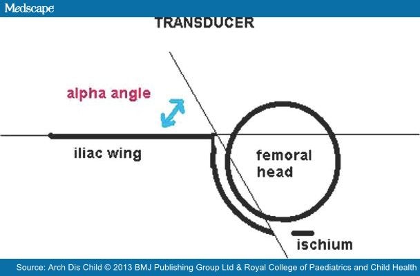
Figure 1.
 
Illustration of the α angle denoting the slope of the bony acetabulum. A modified Graf classification used; Type I (normal) α angle >60°, Type II α angle 43–59°, Type III α angle <43°, Type IV subluxable, dislocated dislocatable hip.
'Pathological' sonographic DDH was considered to be Graf Type III, IV or hip joint irreducibility.[1,5] As 90% of Graf Type II dysplasia spontaneously resolve, the majority of Graf Type II hips were considered to be 'physiological'.[9–11] All Graf Type II hips were followed up until normal or treated if they deteriorated. If the sonographic hip dysplasia deteriorated, the worst classification was recorded in the data analysis (ie, Graf Type III rather than Graf Type II).
Any infant, who presented after the age of 4–6 months with LHA, was initially assessed with a plain pelvic radiograph (although the hip was in addition imaged sonographically if the child was below 40 weeks old). In these cases, the diagnosis of pathological hip dysplasia was made using the radiological maturation curve of Tonnis[12] in which a pathological hip dysplasia was diagnosed if the acetabular index was below two SD of the mean for that age (the lowest 2.5% of the population).
The study was not completed until 4 years after 31 December 2005, in order to identify other cases of irreducible hip dislocation or 'pathological' dysplasia requiring surgery which were born within the 10 years of the study but which presented to the clinic at a later date (ie, 'late' cases).
Statistical analysis was conducted using XLstat comparing the three main groups of patients, namely unilateral LHA, bilateral LHA and no LHA to determine the sensitivity, specificity of the tests as well as the PPV and negative predictive values (NPV) of LHA.

Results

Limited Hip Abduction

A total of 2876 patients were examined clinically and with hip ultrasound imaging over the study period. Of these, 492 patients (17%) had LHA with 2384 patients (83%) being normal with no limitation of hip abduction on the initial clinical assessment. The sonographic hip imaging results are presented in Figure 2.
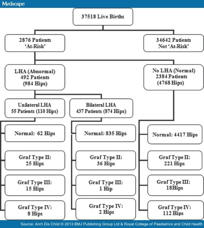
Figure 2.
 
Algorithm of limited hip abduction (LHA) and the ultrasound hip imaging results.
In the total sample of the 'at risk' or unstable hips (5752 hip joints), initial ultrasound showed that 5314 (92.4%) were normal, 282 (4.9%) were Graf Type II, 34 (0.6%) were Graf Type III and 122 (2.1%) were Graf Type IV. Of the 122 Graf Type IV hips (92 patients), 8 (6.6%) were in the unilateral LHA group (all on the clinically abnormal hip), 2 (1.6%) were in the bilateral LHA group (two different patients each having one hip affected) and the remaining 112 (91.8%) in the no LHA group. There were 62 patients with unilateral Graf Type IV and 30 with bilateral Graf Type IV hips (subluxation or dislocation).
The mean age of patients (regardless of the presence or absence of dysplasia and including those presenting late) were unilateral LHA group 131.5 days (95% CI 108.3 to 154.7 days), bilateral LHA group 79.1 days (95% CI 75.6 to 82.5 days) and no LHA group 44.2 days (95% CI 39.9 to 49.7 days). The difference between the ages in the unilateral and bilateral LHA group and the no LHA group was significant (p<0.0001).
If unilateral LHA is only assessed with cases of 'pathological' DDH presenting after the age of 8 weeks (Graf Type III, IV and irreducible hip dislocation), the sensitivity was 78.3%, the specificity was 92.9%, the PPV was 54.7% and the NPV was 97.3%. As the diagnosis of sonographic 'pathological' DDH is likely to have fewer false positive and negative cases, these results are likely be more accurate than if all sonographic dysplasias are assessed in the analysis.
In the no LHA group, there were 82 patients (52 unilateral and 30 bilateral) with 112 Graf Type IV hips (mean age 11.3 days 95% CI±8 days). All the Graf Type IV hips were 'early' dislocatable (within weeks) with only two patients (two hips) requiring later open reduction for failed conservative treatment in a Pavlik harness.
In the bilateral LHA group, there were two patients each presenting with one Graf Type IV hip.
As a clinical screening test the sensitivities, specificities, PPVs and NPVs for unilateral and bilateral LHA as well as the values for unilateral LHA according to age are shown in .

Table 1.  Sensitivity, specificity, positive predictive value (PPV) and negative predictive value (NPV) for unilateral and bilateral LHA and dysplasia of the hip

DysplasiaSensitivity %Specificity %PPV %NPV %
Unilateral LHA
Type III, IV (n=23)
14.499.34097.3
Bilateral LHA
Type III, IV (n=3)
2.241.10.381.8
Unilateral LHA
Type III, IV
Age <8 weeks (n=0)
097.5073.6
Unilateral LHA
Type III, IV
Age >8 weeks (n=23)
78.392.954.797.5
In this 10-year study, the overall dislocation rate requiring open reduction was 0.53–0.59 per 1000 (20 infants/22 hips) and including the surgical treatment of persistent pathological radiological dysplasia the rate was 0.69 per 100 (26 cases). The two bilateral irreducible hip dislocations (1:18 759) both presented after the age of 11 months. Ninety-five per cent of the irreducible hips were identified in females (19 out of 20), whereas in late dysplasia requiring surgery, the 83.34% were in males (5 out of 6). Surprisingly of the 20 irreducible hips, 40% (8 cases) were diagnosed before 3 months (of age), 20% (4 cases) at 3–6 months, 5% (1 case) at 6–12 months and 35% (7 cases) between 12 and 20 months. No cases of irreducible dislocation were diagnosed after 20 months of age.

Discussion

Despite a paucity of evidence supporting its value in improving outcomes, universal clinical screening for DDH, with or without the addition of selective or universal ultrasound hip imaging, remains a well-established method for its detection.[13,14]
Klisic[15] described DDH as a range of hip disorders ranging between dysplasia and irreducible dislocation. This is a dynamic condition in which there can be resolution or deterioration of hip instability or dysplasias. Klisic's[15] definition of DDH includes physiological (or immature) dysplasias (Graf Type II) that generally resolve without treatment. This has resulted in an inconsistency in the literature, as some studies include Graf Type II dysplasias as pathological resulting in high diagnostic and treatment rates of 5–7%. The term DDH does not include neurological, syndromal or neuromuscular cases.[16]
The lack of a practical confirmatory 'gold-standard' clinical or sonographic diagnostic test coupled with a consistent lack of 'known' risk factors in between 69% to 73% of cases makes identification of pathological DDH difficult.[5,17] Clinical examination using provocative tests such as the Ortolani or Barlow manoeuvres continue to be the mainstay of initial diagnosis.[18] These clinical tests are not reliable enough to be considered to be an ideal screening method and should be considered surveillance.[19] Within 1 month of age, 88% of clinically diagnosed neonatal unstable hip joints may spontaneously stabilise.[20] There are very little reliable data on the natural history of Graf Type III hip dysplasia. However, some Graf Type III hips may spontaneously resolve.[9] If this is added to the vague definition of DDH,[18] there is some doubt on what constitutes 'true' pathological DDH. This makes research into this subject difficult and possibly subjective. Some consider the only 'true' pathology in DDH is the rate of irreducible dislocation of the hip.[5] The rate of irreducible hip dislocation in this study is consistent with the best universal clinical or selective screening programmes.[5,21] There is no internationally agreed definition of 'pathological' DDH. However, the general consensus is that in neonatal and infant DDH Graf Type III, IV and irreducible hip dislocation should be treated as pathological.[1,5] Screening for 'pathological' hip dysplasia is not possible as the true incidence or prevalence of this condition in the adult is unknown. The relationship between adult acetabular dysplasia (diagnosed radio logically) and neonatal hip dysplasia (diagnosed sonographically) is unknown. Although up to 30% of total hip replacements are thought to be related to adult hip dysplasia,[22,23] there is no direct proven relationship with neonatal sonographic acetabular abnormalities.
Additional physical signs previously considered to be associated with DDH, including asymmetrical thigh and gluteal skin folds, have little value in diagnosis.[14,15,20,24]
The numbers (%) of 'pathological' sonographic hip dysplasias and/or hip instability from this study are within the accepted range published within the literature.[25] The relationship between LHA and DDH remains controversial. Previous reports have suggested a link with worsening limitation,[4] less severe dysplasia[26] or even dysplasia in the contralateral hip.[27] The definition of LHA may be vague, and like the Ortolani/Barlow manoeuvres, a positive or negative result may suffice in these rather subjective tests. The sonographic hip imaging and clinical hip examinations was undertaken by the same examiner. This increases the likelihood of unintentional bias though this was unavoidable in this study as the clinic was Consultant based (RWP). The senior author, however, has over 20 years experience, and his ultrasonography image quality and interpretation have been independently validated as accurate. Selective 'screening' for DDH (clinical examination combined with hip sonography) is unusual with few orthopaedic practitioners undertaking this in the UK. As in Ortolani/Barlow manoeuvres, the experience of the examiner may be an important factor in the accurate diagnosis of LHA. Irritable infants can make examination difficult or allow for inaccurate recording in inexperienced hands.
While LHA has been identified to be present in DDH, the onset of the sign has not been accurately identified. In our previous study,[1] we noted a low level of LHA in the 'early' dislocatable (5.9%) group compared with those that presented late (87.5%) or early irreducible group (100%). Neonatal laxity is likely to be the reason for the absence of LHA in the neonate.[28] The lack of LHA in the newborn in DDH should be differentiated from the so-called moulded baby syndrome in which LHA is present at birth along with pelvic obliquity, scoliosis, plagiocephaly and postural foot abnormalities. In this condition while LHA is present at birth, there is no association with DDH and ultimately no further treatment is required for the hips.[29,30]
Sixty per cent of the irreducible hip dislocations presented after the age of 3 months. This confirms the failure of universal neonatal and 6-week General Practitioner (GP) clinical screening guidelines in addition to selective at risk sonographic hip screening in identifying and preventing the majority of irreducible hip dislocations.
Our study has shown that the sensitivity of this test was low when all types of sonographic dysplasia are included in the analysis, suggesting that LHA is not exclusive to 'pathological' DDH. Its value, however, markedly increased after the age of 8 weeks, which may also be the time that neonatal laxity itself diminishes. This is 4 weeks earlier than the accepted time frame when LHA becomes reliable.[6] While not commenting upon the precise mode of onset of LHA, other study has reported similar sensitivities of unilateral LHA (69%) in a population of infants over the age of 3 months.[31] Assessing an asymmetrical response to range of movement is easier than a symmetrical one and may account for the very low sensitivity of bilateral LHA and DDH.
We do not consider bilateral LHA in an infant <8 weeks of age to be an accurate clinical sign in the diagnosis of pathological DDH. Bilateral LHA can be rarely associated with bilateral hip dislocations; however, from a cohort of over 37 000 patients over 10 years, we have had only two cases of bilateral hip dislocations both of whom presented late. It is clearly implausible to justify effective clinical screening for such a rare condition. On the rare occasion where a walking infant presents with a severe bilateral LHA in association with postural or gait abnormality (ie, an increased lumbar lordosis and a waddling Trendelenburg gait), it could signify serious hip pathology and a pelvic radiograph should be arranged and assessed.
The high sensitivity and specificity of unilateral LHA after 8 weeks of age suggests that it is an important clinical sign which should be actively sought. This may be particularly true for the secondary general practitioner/health professional (primary healthcare) clinical hip screening programmes which recommend clinical hip screening at 6–8 weeks of age.[18,32] Detection may allow earlier intervention and potentially reduce the rate of 'late' irreducible hip dislocation presentation. Training into the importance of unilateral LHA presenting after the age of 6–8 weeks of age should be emphasised. Ideally, the clinical examination should be undertaken by small groups of experienced clinicians as wide groups of inexperienced clinicians may miss this important clinical sign. Such experienced small groups have been shown to improve effectiveness of accurate diagnosis in neonatal hip instability in Sweden.[33]
The current recommended 6-week assessment may be too early and may miss the development of the LHA. Delaying the assessment until 8 weeks may be advantageous. This change would require a proper multicentre controlled comparative trial between the different timings (6 weeks vs 8–12 weeks), showing a clear advantage before any change in timing could be recommended. Due to the low incidence of 'pathological' DDH, this series would take many years and would be difficult to organise.
In conclusion, this study identifies a time-dependent association with unilateral LHA in the diagnosis of 'pathological' DDH after the age of 8 weeks. The presence of bilateral LHA in the young infant may be a normal variant in the majority and is a poor screening sign for determining the presence of pathological DDH due to its low sensitivity and poor PPV. LHA should be actively sought after 8 weeks of age and if present should prompt for referral to a specialist (Paediatric Orthopaedic Surgeon or expert hip sonographer) followed by a formal ultrasound or radiographic examination to confirm whether or not the hip is developing in a satisfactory manner.

Sidebar 1

What Is Known About This Topic

  • The lack of a practical confirmatory 'gold-standard' clinical or sonographic makes identification of developmental dysplasia of the hip (DDH) difficult.
  • The relationship of limitation of hip abduction (LHA) and DDH remains controversial. While LHA has been identified to be present in DDH, the onset of the sign has not been accurately identified.

Sidebar 2

What This Study Adds

  • The bilateral LHA sign may be a normal variant in infants and has a low sensitivity and positive predictive value.
  • Our study shows the high sensitivity and specificity of unilateral LHA after 8 weeks of age suggesting that it is an important clinical sign which should be actively sought.

References

  1. Jari S, Paton RW, Srinivasan MS. Unilateral limitation of abduction of the hip. A valuable clinical sign for DDH? J Bone Joint Surg[Br] 2002;84B:104–7.
  2. Godward S, Dezateux C. Surgery of congenital dislocation of the hip in the UK as a measure of outcome of screening. MRC Working Party on Congenital Dislocation of the Hip. Medical Research Council. Lancet 1998;351:1149–52.
  3. Terjesan T. ULtrasound as the primary imaging method in the diagnosis of hip dysplasia in children aged <2 years. J Pediatr Orthop 1996;5:123–8.
  4. Stoffelen D, Urlus M, Molenaers G, et al. Ultrasound, radiographs and clinical symptoms in developmental dislocation of the hip: a study of 170 patients. J Pediatr Orthop B 1995;4:194–9.
  5. Paton RW, Hinduja K, Thomas CD. The significance of at-risk factors in ultrasound surveillance of developmental dysplasia of the hip. J Bone Joint Surg [Br]2005;87B:1264–6.
  6. Homer CJ, Balz RD, Hickson GB, et al. American Academy of Pediatrics, Clinical Practice Guideline: Early Detection of Developmental Dysplasia of the Hip. Committee on Quality Improvement, Subcommittee on Developmental Dysplasia of the Hip. Pediatrics 2000;105:896–905.
  7. Graf R, Tschauner C, Klapsch W. Progress in prevention of late developmental dislocation of the hip by sonographic newborn hip screening—results of a comparative follow-up-study. J Pediatr Orthop B 1993;2:115–21.
  8. Harcke HT, Clarke NM, Lee MS, et al. Examination of the infant hip with real time ultrasonography. J Ultrasound Med 1984;4:131–7.
  9. Castelein RM, Dauter AJ, deVlieger M, et al. Natural history of ultrasound hip abnormalities in clinically normal newborns. J Pediatr Orthop 1992;12:423–7.
  10. Wood MK, Conboy V, Benson MKD. Does early treatment by abduction splintage improve the development of dysplasic but stable hips? J Pediatr Orthop2000;20:302–5.
  11. Rosendahl K, Markestad T, Lie RT. Developmental dysplasia of the hip. A population based comparison of ultrasound and clinical findings. Acta Paediatr1996;85:64–9.
  12. Tonnis D. Normal value of the hip joint for the evaluation of X-rays in children and adults. Clin Orthop Relat Res 1976;119:39–47.
  13. Patel H. Preventative health care: 2001 update: screening and management of developmental dysplasia of the hip in newborns. CMAJ 2001;164:1667–77.
  14. Shipman SA, Hefand M, Moyer VA, et al. Screening for developmental dysplasia of the hip: as systemic literature review for the US Preventative Services Task Force. Pediatrics 2006;117:e557–76.
  15. Klisic PJ. Congenital dislocation of the hip—a misleading term: brief report. J Bone Joint Surg 1989;71:136.
  16. Bialik V, Bialik GM, Blazer S, et al. Developmental dysplasia of the hip: a new approach to incidence. Pediatrics 1999;103:93–9.
  17. Bache CE, Clegg J, Herron M. Risk factors for developmental dysplasia of the hip: ultrasonographic findings in the neonatal period. J Pediatr Orthop B2002;11:212–18.
  18. Standing Medical Advisory Committee (SMAC). Screening for the detection of congenital dislocation of the hip. London: Department of Health and Social Security, 1986.
  19. Jones D. Neonatal detection of developmental dysplasia of the hip. J Bone Joint Surg [Br] 1998;80:943–5.
  20. Barlow T. Early diagnosis and treatment of congenital dislocation of the hip. J Bone Joint Surg [Br] 1962;44-B:292–301.
  21. Macnicol MF. Results of a 25 year screening programme for neonatal hip instability. J Bone Joint Surg [Br] 1990;72:1057–60.
  22. Editorial. Screening for congenital hip dysplasia. Lancet 1991;337:947–8.
  23. Furnes O, Lie SA, Espehaug B, et al. Hip disease and the prognosis of total hip replacements. A review of 53,698 primary hip replacements reported to the Norwegian Arthroplasty Register 1987–99. J Bone Joint Surg [Br] 2001;83:579–86.
  24. Palmén K. Preluxation of the hip joint: diagnosis and treatment in the newborn and the diagnosis of congenital dislocation of the hip joint in Sweden during the years 1948–1960. Acta Paediatr 1961;50(Suppl 129):1–71.
  25. Rosendahl K, Markestad T, Lie RT. Developmental dysplasia of the hip: prevalence based on ultrasound diagnosis. Pediatr Radiol 1996;26:635–9.
  26. Bialik V, Wiener F. Sonography of suspected developmental dysplasia of the hip: a description of 3624 hips. J Pediatr Orthop 1993;2:152–5.
  27. Green NE, Griffin PP. Hip dysplasia associated with abduction contracture of the contralateral hip. J Bone Joint Surg [Am] 1982;64:1273–81.
  28. Wynne-Davies R. Acetabular dysplasia and familial joint laxity: Two etiological factors in congenital dislocation of the hip. J Bone Joint Surg 1970;52-B:704–16.
  29. Good C, Walker G. The hip in the moulded baby syndrome. J Bone Joint Surg [Br] 1984;66:491–2.
  30. Buxton RA, Macnicol MF. Infantile skeletal skew: the use of ultrasound in management. J Pediatr Orthop B 2004;13:75–80.
  31. Castelein RM, Korte J. Limited hip abduction in the infant. J Pediatr Orthop 2001;21:668–70.
  32. Newborn and Infant Physical Examination (NIPE). http:/nipe.screening.nhs.uk
  33. Duppe H, Danielsson LG. Screening of neonatal instability and of developmental dislocation of the hip. A Survey of 132,601 Living newborn infants between 1956 & 1999. J Bone Joint Surg [Br] 2002;84-B:878–85.