Παρασκευή 31 Μαΐου 2013

Guidance for Identifying Motor Delays in Children


Guidance for Identifying Motor Delays in Children

Diedtra Henderson
May 30, 2013

An algorithm that includes formal developmental screening of children for possible motor delays and variations in muscle tone at their 9-, 18-, 30-, and 48-month well-child visits could help affected children receive diagnostic evaluations and treatments in a more timely fashion, according to new guidelines.
Garey H. Noritz, MD, from the Council on Children With Disabilities, and coauthors, including members of the Neuromotor Screening Expert Panel, present guidance for pediatric primary care providers in an article published online May 27 in Pediatrics.
Gross motor delays are common among children, according to the authors. About 3.3 children per 1000 have more permanent motor disabilities, such as cerebral palsy, and 6% of children have developmental coordination disorder. According to the multidisciplinary expert panel, early diagnosis can help reduce family stress, and earlier access to available treatments can improve outcomes for children with neuromuscular diseases. The most frequently used developmental screening instruments, however, have not been validated on children with motor delays.
During focus groups involving 48 pediatricians attending an American Academy of Pediatrics conference in 2010, practitioners reported uncertainty in detecting and diagnosing motor delays in children.
Underscoring recommendations made by the academy in 2006, the expert panel now recommends using a standardized test to provide children with periodic developmental screening during their 9-, 18-, and 30-month well-child visits. By 9 months, an infant should be able to roll to both sides and sit without help. By 18 months, toddlers should have mastered sitting, standing, and walking on their own. Although most motor delays would have already been evident at a younger age, by 30 months, more subtle impairments may become evident, and progressive neuromuscular disorders may begin to appear. By 48 months, preschoolers should climb stairs without help, skip on 1 foot, feed themselves, and demonstrate such fine motor milestones as drawing stick people and fastening medium-sized buttons.
Pediatricians not only should watch how children perform a requested task but also should pay keen attention to their general posture, play, and spontaneous motor functions, the authors counsel.
Muscle tone can provide an additional clue of a possible neuromotor delay, such as cerebral palsy. A magnetic resonance imaging scan can be ordered for patients with heightened muscle tone, and serum creatine kinase concentrations should be measured in children with decreased muscle tone. Such interventions can occur at the same time children are referred to specialists for diagnosis.
Pediatricians treating a child with mild abnormalities should develop a time-definite follow-up plan should they worsen or develop new symptoms, such as regression in motor skills or loss of strength. Acting quickly on such clinical changes can help expedite appropriate medical attention.
"The initial responsibility for identifying a child with motor delay rests with the medical home. By using the algorithm presented here, the medical home provider can begin the diagnostic process and make referrals as appropriate," the authors conclude.
Financial support for the clinical report was provided by the American Academy of Pediatrics. The authors have disclosed no relevant financial relationships.
Pediatrics. Published online May 27, 2013. Abstract

Τετάρτη 29 Μαΐου 2013

How to Evaluate Muscle Weakness in Infants and Children


How to Evaluate Muscle Weakness in Infants and Children

Mark E. Swanson, MD, MPH
Dec 10, 2012

Neuromuscular diseases, such as muscular dystrophy, are individually rare. Yet, pediatric clinicians frequently encounter children with motor delay, and an approach to identify serious causes of such delays is needed. The average age at diagnosis of Duchenne muscular dystrophy is approximately 5 years, although parents recognize the earliest symptoms when their child is a mean 2.5 years of age.[1] This diagnostic delay has not improved in 2 decades, and such a delay hinders access to information about care options, relevant clinical trials, and support networks for the child's disorder. Delays in diagnosis also impede access to early intervention and other healthcare services. On the other hand, early diagnosis facilitates the family's access to genetic counseling to learn about family planning options. Family stress can be significant when an accurate diagnosis of a serious neuromuscular disorder is delayed. Families typically see several clinicians and the child undergoes unnecessary testing before the child is referred to a specialist familiar with neuromuscular disorders.
The Centers for Disease Control and Prevention (CDC), in collaboration with the National Task Force for Early Identification of Childhood Neuromuscular Disorders, has developed a new Web-based diagnostic tool, the Surveillance and Referral Aid for Primary Care Providers to assist providers in primary care, rehabilitation medicine, and physical and occupational therapy in the evaluation of children with motor delay, including identification of early signs and symptoms of neuromuscular disorders.
The Website of the National Task Force for Early Identification of Childhood Neuromuscular Disorders, ChildMuscleWeakness.org, focuses on evaluation of children 6 months to 5 years of age. Neuromuscular diseases that might present in this age range include (but are not limited to) spinal muscular atrophy types 2 and 3, congenital muscular dystrophies, limb-girdle muscular dystrophy, Charcot-Marie-Tooth disease, some presentations of myotonic muscular dystrophy, Duchenne and Becker muscular dystrophy, and metabolic muscle diseases.
Figure. A child with spinal muscular atrophy type 2, at 2 years of age.
ChildMuscleWeakness.org provides guidance on motor surveillance and screening that expands on the Bright Futures guidelines developed by the American Academy of Pediatrics. The surveillance guidance includes an aid to the assessment of motor development milestones, "signs of weakness by age," including recommendations for evaluating the following milestones:
  • Infant+: head lag on pull to sit;
  • Age 6+ months: achieving and maintaining sitting; and
  • Age 12+ months: rising to stand from the floor and gait (walking and running)
If a delay is found using the surveillance aid, an accompanying motor delay algorithm provides guidance on testing and referral. Clinical pearls outline red flags that indicate the need for an urgent referral to a neurologist:
  • Tongue fasciculations;
  • Loss of motor milestones; or
  • Creatine phosphokinase (CK) level > 3 times normal (however, children with some neuromuscular disorders have normal CK levels).
Many neuromuscular conditions increase the risk for malignant hyperthermia with anesthesia use, and anticipated surgery should increase the urgency of a diagnostic evaluation. The core tools found on ChildMuscleWeakness.org have been endorsed by the American Academy of Pediatrics.
ChildMuscleWeakness.org is designed for clinicians and includes many resources and tools, such as an extensive library of videos showing examples of early and later signs of muscle weakness compared with normal motor development. Information is presented on how to differentiate between muscle weakness caused by diseases of the brain (central causes, such as cerebral palsy) and neuromuscular diseases (peripheral causes, such as Duchenne muscular dystrophy). Other resources include guidance on communicating about motor concerns with families, case studies, teaching tools, and a summary of terms parents use to describe motor delays and weakness. The site highlights the importance of CK testing when motor delay is identified and the cause of the delay is not clear.
The contents of the ChildMuscleWeakness.org Website are the sole responsibility of the National Task Force for Early Identification of Childhood Neuromuscular Disorders and do not necessarily represent the official views of the Centers for Disease Control and Prevention. ChildMuscleWeakness.org was developed under CDC cooperative agreement #DD08-805.
Web Resource
Mark Swanson, MD, MPH, is Senior Medical Adviser to the Division of Human Development and Disability of the National Center on Birth Defects and Developmental Disabilities, Centers for Disease Control and Prevention.
A graduate of Princeton University, the University of Colorado School of Medicine, and the University of North Carolina School of Public Health, Dr. Swanson has been board-certified in Pediatrics and Developmental-Behavioral Pediatrics during his 35-year career. He has extensive experience in program development, clinical services, research, and training in community and university settings. He was the first Director of Partners for Inclusive Communities at Arkansas' University Center on Disabilities and oversaw its growth from 1990 to 2005 before joining the Centers for Disease Control and Prevention in the Division of Human Development and Disability (DHDD). For 3 years, he has directed a multifaceted program that includes state-level health promotion programs for persons with disabilities, extramural research projects, the National Spina Bifida Program, information resource centers addressing physical activity, intellectual disability, paralysis and limb loss, and an intramural research program focusing on health disparities in persons with disabilities. Swanson currently serves as Senior Medical Adviser in DHDD, which has additional programs in ADHD, Tourette syndrome, muscular dystrophy, fragile X syndrome, and hearing loss. He has a career-long commitment to full inclusion and participation of persons with disabilities in all aspects of life.

References

  1. Ciafaloni E, Fox DJ, Pandya S, at al. Delayed diagnosis in Duchenne muscular dystrophy: data from the Muscular Dystrophy Surveillance, Tracking, and Research Network (MD STARnet). J Pediatr. 2009;155:380-385. Abstract

New Tool Aids in Diagnosis of Pediatric Migraine


New Tool Aids in Diagnosis of Pediatric Migraine

Fran Lowry
Jan 17, 2013

A new structured interview has been shown to be a reliable and valid method for detecting migraine in children and teens in both clinical and community settings.
The Diagnostic Interview of Headache Syndromes–Child Version (DIHS-C) can be administered by nonclinicians to detect criteria for headache disorders, as set out in the second edition of the International Classification of Headache Disorders among children aged 7 to 18 years, write Tarannum Lateef, MD, MPH, from the National Institute of Mental Health, Bethesda, Maryland, and her colleagues.
Compared with an expert neurologist's diagnosis of migraine, the sensitivity with use of this tool was 98%. "In other words, we were able to accurately diagnose migraine in 98% of the children who had it with the interview," Dr. Lateef told Medscape Medical News.
Their findings were published online in Pediatrics.
Common and Debilitating
"Childhood headache is quite common and in many cases can even be debilitating," Dr. Lateef said. "We wanted to develop a tool that would allow for early and accurate diagnoses of headache syndromes in childhood. This is the best way to ensure appropriate treatment and improve outcomes for affected youth."
To this end, Dr. Lateef and her group conducted 104 pediatric headache interviews (53 in boys, 51 in girls) that were done as part of a community-based family study of migraine. In 40 of the children, a clinician had previously diagnosed migraine.
The researchers then compared the diagnosis of pediatric migraine made by the interview with the diagnosis made by the study neurologists.
They found that the DIHS-C was a reliable and valid means for ascertaining migraine in both clinical and community settings.
The overall sensitivity of the interview diagnosis compared with the expert neurologists' diagnosis of migraine was 98%, and specificity was 61%.
Dr. Lateef suggested that doctors can use this tool in their offices as an initial history-gathering method administered by a nonphysician. "The treating physician can then use the information. Often diagnoses may be overlooked since the physician does not have enough time to go over an extensive interview process for various diagnoses, and this interview can save time."
She added that she and her colleagues would like to see increased recognition of pediatric migraine in both community and clinical settings. "We hope that use of this diagnostic interview will lead to better management and better outcomes for migraine sufferers starting at an early age."
Migraine a Pediatric Problem
Commenting on this study for Medscape Medical News, Paul Graham Fisher, MD, the Beirne Family Professor of Pediatric Neuro-Oncology and professor, neurology and pediatrics, at Stanford University and Lucile Packard Children's Hospital, Palo Alto, California, noted, "Lateef and her team showed nicely that a structured interview, performed by nonphysician personnel, could detect very well those children and teens suffering from headache."
He noted limitations of the study, including that the small sample was "enriched" with migraine patients, and that the tool is long.
Despite these issues, "the work demonstrates further that migraine is indeed a pediatric problem and can be screened for by primary care personnel, not just pediatricians and specialists.
"While the researchers' tool might help best in the investigational arena, their approach, and perhaps that of successors, offers hope that more children who suffer from migraine can be identified in everyday clinics and remedied, so they can continue on with other aspects of their lives," Dr. Fisher concluded.
The study was supported by the National Institutes of Health. Dr. Lateef and Dr. Fisher have disclosed no relevant financial relationships.
Pediatrics. 2013;131:e96-e102. Published online December 24, 2012. Abstract

Most Common Foods for Foodborne Illness: CDC Report


Most Common Foods for Foodborne Illness: CDC Report

Kathleen Doheny
Jan 30, 2013

Every year, about 48 million Americans — 1 in 6 — get a food-borne illness. 
Often, the culprit food is a mystery.
Now, a new CDC report based on a decade of data offers the first comprehensive estimates of which foods are to blame.
Produce accounts for nearly half of the illnesses, and the norovirus is often to blame. Norovirus causes about 20 million cases of "stomach flu" each year. A new strain is going around the U.S.
Poultry is the food source with the most fatal infections. Salmonella and listeria are the common germs causing these infections.
"There is food-borne illness caused by a wide variety of foods," says researcher John Painter, DVM, an epidemiologist at the CDC.
"We didn't attempt to assign any risk for [specific] foods in the study," he says. The report does not mean people should avoid any foods, especially healthy choices such as produce, he says. The foods most often involved in outbreaks are often the foods we eat frequently and are part of a healthy diet.
The report is published in the journal Emerging Infectious Diseases.
According to the CDC, the report is the most complete effort to attribute illness to specific foods.
Food-borne Illness Report: Details
Food-borne illness sends 128,000 Americans to the hospital each year and kills 3,000 annually.
To compile the report, the CDC evaluated nearly 4,600 food-borne disease outbreaks from 1998 to 2008. The researchers had information on both the specific food causing the outbreak and the specific type of illness.
They estimated how much food-borne illness is accounted for in 17 food categories. Among the findings:
  • Nearly half of illnesses were linked to produce. Produce includes fruits, nuts, leafy greens, and other vegetables.
  • Of these produce foods, leafy greens were most often involved in food-borne illness. Norovirus was often the germ involved.
  • Dairy was the second most frequent food source for infections.
  • Contaminated poultry was to blame for the most deaths, involved in 19% of fatal cases. Many were linked to listeria and salmonella infections. Together, meat and poultry were to blame for 22% of illnesses and 29% of deaths.
  • All 17 food categories were involved in some outbreaks, but the frequency for each category varied.
  • Half of the outbreaks with a known food involved foods with ingredients from several food categories.
  • Dairy and eggs accounted for 20% of illnesses and 15% of deaths.
  • Fish and shellfish accounted for 6.1% of illnesses and 6.4% of deaths.
Food handlers are often to blame for norovirus outbreaks, according to a previous CDC study.
This virus is in the vomit and stool of infected people. It can be passed when infected food handlers don't wash their hands.
Preventing Food-borne Illness
Simple steps, both at home and when eating out, can lower your risk of food-borne illness.
"Being careful of cross-contamination in the kitchen is one of the most important elements," Painter says.
For instance, slicing chicken on a cutting board, then using it to prepare a salad, raises infection risk.
Wash hands often, especially when preparing food, to cut down the risk of transmitting norovirus, Painter says.
Don Schaffner, PhD, a food microbiologist at Rutgers University who was not involved in the report, recommends that people wash raw food. "If you are buying your own produce, such as head lettuce, you want to wash it properly in your kitchen," he says.
However, if you buy the packaged, triple-washed products, he recommends against washing it again. The risk of contamination in your kitchen has been found to outweigh the risk of triple-washed lettuce making you sick.
Home cooks just getting over a bout of vomiting and diarrhea should not be handling food, Schaffner says.
When eating out, if you notice employees not washing their hands, report that to the manager or your local health department, Painter says. If many people get sick eating out after having the same meal, call the county health department.
"It's those outbreaks that are the basis of outbreak reports," he says.
Schaffner reports serving as a consultant to the food industry and for companies making hand sanitizer and antibacterial soaps.
SOURCES:
Painter, J. Emerging Infectious Diseases, March 2013.
John Painter, DVM, epidemiologist, CDC.
Don Schaffner, PhD, professor of food microbiology, Rutgers University; science communicator, Institute of Food Technologists.

A New Technique for Fast and Safe Collection of Urine in Newborns


A New Technique for Fast and Safe Collection of Urine in Newborns

María Luisa Herreros Fernández, Noelia González Merino, Alfredo Tagarro García, Beatriz Pérez Seoane, María de la Serna Martínez, María Teresa Contreras Abad, Araceli García-Pose,
Arch Dis Child. 2013;98(1):27-29. 

Abstract and Introduction

Abstract

Aim To describe and test a new technique to obtain midstream urine samples in newborns.
Design and methods This was a prospective feasibility and safety study conducted in the neonatal unit of University Infanta Sofía Hospital, Madrid. A new technique based on bladder and lumbar stimulation manoeuvres was tested over a period of 4 months in 80 admitted patients aged less than 30 days. The main variable was the success rate in obtaining a midstream urine sample within 5 min. Secondary variables were time to obtain the sample and complications.
Results This technique was successful in 86.3% of infants. Median time to sample collection was 45 s (IQR 30). No complications other than controlled crying were observed.
Conclusions A new, quick and safe technique with a high success rate is described, whereby the discomfort and waste of time usually associated with bag collection methods can be avoided.

Introduction

Clean urine samples are necessary to accurately diagnose several diseases in infants, especially urinary tract infections (UTIs). A wide range of clinical interventions for urine collection is described in the literature, including non-invasive and invasive methods. The most common non-invasive technique is urine collection using sterile bags, which is associated with significant patient discomfort and contamination of samples. Obtaining a clean-catch urine sample is the recommended method for urine collection in children able to co-operate. However, in children lacking sphincter control, urine catch is more difficult and time-consuming and invasive methods (catheterisation and needle aspiration of urine from the bladder) are sometimes needed.[1,2]
There are some stimulation techniques that facilitate emptying of the bladder in situations of bladder dysfunction.[3] We hypothesised that the use of such methods in newborns could facilitate the collection of a clean-catch urine sample.
The aim of this study was to determine the success rate and safety of a new non-invasive technique to obtain clean-catch urine samples in newborns.

Methods

This was a prospective feasibility and safety study conducted in Infanta Sofía University Hospital, Madrid (a secondary centre, with a 16-bed neonatal unit, a 30-bed nursery unit and a 24-bed paediatric ward). The study was carried out over 4 months (January–April 2010). Patients consisted of 90 consecutively admitted infants aged under 30 days who needed a urine analysis according to their attending physician. Exclusion criteria were: (1) poor feeding, (2) dehydration, (3) need for an immediate sterile urine sample obtained by an invasive method or a clinical situation that did not allow time to obtain a midstream urine sample, (4) any medical condition that ruled out manipulation, and (5) drug administration prior to urine collection.
The clinical research ethics committee of La Paz University Hospital, Madrid, Spain, approved this study.

Technique

Two people (trained nurses and/or physicians) were needed to perform the procedure, and a third to measure the time taken. This technique involves a combination of fluid intake and non-invasive bladder stimulation manoeuvres.
The first step is either breast-feeding or providing formula intake appropriate to the age and weight of the newborn. In babies fed infant formula, 10 ml was provided on the first day of life, increasing to 10ml per day during the first week. From the second week onwards, 25 ml/kg were administered before the onset of stimulation. Twenty-five minutes after feeding, the infant's genitals were cleaned thoroughly with warm water and soap and dried with sterile gauze. A sterile collector was placed near the baby in order to avoid losing urine samples. Before performing the technique, we administered non-pharmacological analgesia, such as non-nutritive sucking or 2% sucrose syrup, to prevent/lessen crying.
The second step is to hold the baby under their armpits with their legs dangling. One examiner then starts bladder stimulation which consists of a gentle tapping in the suprapubic area at a frequency of 100 taps or blows per minute for 30 s.
The third step is stimulation of the lumbar paravertebral zone in the lower back with a light circular massage for 30 s.
Both stimulation manoeuvres are repeated until micturition starts, and a midstream urine sample can be caught in a sterile collector (figure 1). Success is defined as the collection of a sample within 5 min of starting the stimulation manoeuvres.
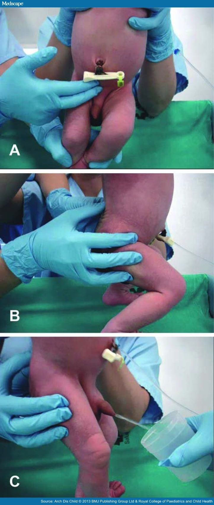
Figure 1.
 
New stimulation technique to obtain midstream urine in newborns. (A) Tapping in the suprapubic area. (B) Stimulation of the lower back. (C) Midstream urine sample collection in a sterile container.

Variables

The main variable was the success rate in obtaining a midstream urine sample within 5 min. Secondary variables were the time taken to obtain the sample and complications. The sample collection time was defined as the time from the beginning of the stimulation procedure (ie, the tapping) to the beginning of sample collection.

Analysis

We analysed the percentage of attempts where urine was obtained ('success'), the time to obtain the sample from the onset of bladder stimulation, age and sex. Categorical variables ('success', sex) are expressed as rates (%) and measurable variables (age, time) as mean (SD), 95% CI, median and IQR. Categorical variables were compared using Pearson's χ2 or Fisher's exact test. Quantitative variables were compared using Student's t test or by the Mann–Whitney U test. All statistical analyses were performed with SPSS software (V.17.0). We assumed significance at the 5% level (p<0.05).

Results

Ninety patients were eligible for the study. Indications for urine collection were neonatal jaundice (49), screening for infection (20), cytomegalovirus screening in low birthweight newborns (14) and renal pelvis dilatation (7). Ten patients were later excluded due to low oral intake leaving 31 girls and 49 boys. Their mean ages were 6.66 days for boys and 6.23 days for girls. There was an 86% success rate (n=69/80).
The mean time for sample collection was 57 s (SD 48.6), median 45 s and IQR 30 s. The mean time spent collecting the sample in males was 60.48 s, median 55 s and IQR 30 s. For females, the mean time was 52.04 s, median 30 s and IQR 30 s. Urine was sometimes obtained before the end of the first cycle of stimulation (<60 s).
No statistically representative differences with regard to sex were found in success rate, time of sample collection or complications. There were no complications, but controlled crying occurred in 100% of infants.

Discussion

We report a new technique to obtain a midstream clean urine sample in newborns, which has a high success rate and a mean time for passing urine of less than 1 min.
Midstream urine collection is the preferred method for adults and older children and is suitable for diagnosing UTI.[1] The collection of spontaneous urine in infants is also useful for UTI investigation, but is a time-consuming and unpredictable task that requires prolonged attention and patience, and so is not widely performed.
The advantages of midstream urine collection and the difficulties performing it in newborns encouraged us to develop a suitable technique for this age group. Invasive methods for obtaining clean urine (suprapubic aspiration and bladder catheterisation) are aggressive and have a high failure rate in newborns due to their anatomical characteristics and irregular voiding pattern.
We based the procedure on manoeuvres described for patients with bladder dysfunction to stimulate bladder emptying through reflex contraction of the detrusor muscle.[3] The detrusor muscle is innervated by the parasympathetic pelvic nerves (S2–S4). The spinal micturition reflex is a simple arch reflex. Distended bladder walls stimulate efferent fibres going to the medulla, the arch reflex is produced in S2–S4, and afferent fibres stimulate the detrusor muscle which contracts to pass urine. This reflex is voluntarily inhibited and controlled in continent individuals by the cortex, but not in newborns. In neonates, it can be triggered, as we propose.
We designed a stimulation technique and protocol and performed a study after nurses and physicians had been trained. We have demonstrated that this technique is effective in obtaining a urine sample in a majority of patients in an easy, safe and fast way. Bag changes, long waiting times and invasive techniques were avoided.
Limitations of this study include the lack of control group. Nevertheless, the time to obtain urine in other published series is much longer.[4] Several factors may have influenced the efficiency of our technique: trained staff performed the procedure and a standardised fluid intake favoured the onset of urination after 20–30 min in this age group.[5]
As far as we know, there is no other standardised method to facilitate micturition in infants. This technique has replaced the use of collection bags as a routine method for newborns in our centre. Other invasive and aggressive methods carrying some risk (suprapubic aspiration or bladder catheterisations) were also avoided.

Conclusions

A new method to obtain midstream urine in newborns is described. It consists of feeding, bladder stimulation and paravertebral lumbar massage. The technique has been demonstrated to be safe, quick and effective. The discomfort and waste of time usually associated with bag collection methods can be avoided, as well as invasive techniques.

Sidebar 1


Vaccination Choice Influenced by Social Networks


Vaccination Choice Influenced by Social Networks

Lara C. Pullen, PhD
Apr 15, 2013

Parents make vaccination decisions largely based on their social networks. In particular, nonconformity recommendations (recommendations not to vaccinate) within the social network were more predictive of vaccination decisions than parents' own perceptions of vaccinations.
Emily K. Brunson, PhD, MPH, from Texas State University in San Marcos, presented the results of her survey in an article published online April 15 in Pediatrics. Dr. Brunson surveyed United States–born, first-time parents who had children aged 18 months or younger and resided in King County, Washington. A total of 126 participants conformed to vaccination recommendations and 70 did not.
In both groups, 95% or more of parents reported having "people networks" (people that they go to for information). Nonconformers, however, were significantly more likely to report "source networks" (sources parents go to for information and advice; 100% versus 80%; P < .001).
Parents undergo a complex process when deciding whether or not to immunize their children. Research in the 1950s first demonstrated how strong social influence can be in guiding personal judgement.
The current study connects immunization decision-making with the pressure to conform to group opinion and explores whether parents choose a social group that reflects their belief and actions or, rather, let their social group dictate their beliefs and actions. Dr. Brunson's data suggest that the social groups dictate the decisions.
The study also allows for the likelihood that social networks sustain and strengthen a vaccination decision. The study reinforces the theory that healthcare decisions in general, and vaccination decisions in particular, are made within a social context.
"Although the provider-parent conversation about vaccines may be the most influential to parent decision-making, these conversations can be difficult to navigate, especially during the increasingly time-limited provider-parent encounter," write Douglas J. Opel, MD, MPH, and Edgar K. Marcuse, MD, MPH, both from the University of Washington in Seattle, in an accompanying editorial. "Public health interventions aimed at shaping the social milieu of immunization decision-making outside the examination room can complement providers' efforts to achieve the full benefit of immunizations within. Our emerging understanding of vaccine decision-making suggests that this priming of the pump is just what is needed."
The author and editorialists have disclosed no relevant financial relationships.

Longer Breast-feeding May Mean Lower Iron Levels


Longer Breast-feeding May Mean Lower Iron Levels

Norra MacReady
Apr 17, 2013

Breast may be best, but relying on it too long may set a child up for iron deficiency later, a new study suggests.
In a cross-sectional analysis of 1647 children aged 1 to 6 years, the odds of iron deficiency increased by 4.8% for each month of breast-feeding, lead author Jonathon L. Maguire, MD, and colleagues report in an article published online April 15 in Pediatrics.
Dr. Maguire, from the Department of Pediatrics and the Li Ka Shing Knowledge Institute of St. Michael’s Hospital; the Pediatrics Outcomes Research Team, Division of Paediatric Medicine, Department of Paediatrics, The Hospital for Sick Children; and the Department of Pediatrics, Faculty of Medicine, University of Toronto, Ontario, Canada, and coauthors studied children visiting 7 primary care clinics participating in The Applied Research Group for Kids (TARGet Kids!) study, which focuses on the long-term health and development of Canadian infants and preschoolers, between December 2008 and June 2011.
Iron stores, as measured by serum ferritin, were the primary outcome measure, with deficiency defined as anything below 14 μg/L. Iron deficiency anemia was defined as a hemoglobin level of 110 g/L or less in the presence of iron deficiency. Breast-feeding duration was determined through maternal recall. Excluded from the study were children with any chronic condition other than asthma and those with evidence of inflammation, as indicated by a C-reactive protein level of 10 mg/L or more, because of concerns that an acute illness might falsely elevate serum ferritin levels.
The analysis was adjusted for potentially confounding covariates including age, sex, birth weight, body mass index, ethnicity, household income, day care attendance, age of introduction to cow's milk or solid food, and current intake of cow's milk.
The study participants had a median age of 36 months (range, 12 - 72 months). Ninety-three percent had been breast-fed, for a median duration of 10 months (range, 0 - 48 months). Their median serum ferritin level was 32 μg/L (range, 2 - 172 μg/L).
The prevalence of iron deficiency and iron deficiency anemia was 9% and 1.5%, respectively. On univariate analysis, every additional month of breast-feeding was associated with a decrease in serum ferritin of 0.21 μg/L (P = .0006), an association that remained significant even after adjustment for the covariates. Younger age, higher birth weight, and greater consumption of cow's milk also were independently associated with lower serum ferritin levels. On secondary analysis, there was a statistically significant association between breast-feeding duration and the odds of iron deficiency (odds ratio [OR], 1.026; 95% confidence interval [CI], 1.004 - 1.050). A total breast-feeding duration longer than 12 months was associated with an adjusted OR for iron deficiency of 1.71 (95% CI, 1.05 - 2.79) compared with a duration of less than 12 months.
The adjusted OR for iron deficiency was 1.048 for each additional month of breast-feeding, or an odds increase of 4.8% per month (95% CI, 1.02 - 1.08).
Potential study limitations include the relatively low prevalence of iron deficiency and anemia and the use of parental self-report to estimate duration of breast-feeding.
At least one outside observer believes these data should not be interpreted to mean mothers should curtail their breast-feeding. "Mothers should exclusively breast-feed for the first 6 months," and then introduce iron-fortified foods while continuing to breast-feed for 1 year or more, Diane Spatz, PhD, RN, director of the Lactation Program at Children's Hospital of Philadelphia in Pennsylvania, told Medscape Medical News. To determine further relationships between breast-feeding and iron status, "it would be important to study the infant's other dietary intake after 6 months."
Still, the authors warn, "no previous study has documented a relationship between breastfeeding beyond 12 months of age and reduced iron stores."
This study was supported by a grant from the Canadian Institutes of Health Research, the Sick Kids Foundation, and St. Michael's Foundation. One study author is a consultant to AstraZeneca, Bristol-Myers Squibb, GlaxoSmithKline, Hoffmann LaRoche, Novartis, and Pfizer. The other authors and Dr. Spatz have disclosed no relevant financial relationships.

Death From Infant Sleep Positioners


Death From Infant Sleep Positioners

William T. Basco, Jr., MD, MS
May 01, 2013

Suffocation Deaths Associated With Use of Infant Sleep Positioners -- United States, 1997-2011

Centers for Disease Control and Prevention

MMWR Morb Mortal Wkly Rep. 2012;46:933-937

Infant Sleep Positioners

Infant sleep positioners are marketed to the public with several health claims, including an overall improvement in sleep quality as well as prevention of sudden infant death syndrome (SIDS). Few of these devices have US Food and Drug Administration (FDA) approval, and those that do are designed for the prevention of gastroesophageal reflux or for the treatment of plagiocephaly. None has a specific indication for prevention of SIDS.

Study Summary

This article summarizes 13 cases of suffocation associated with infant sleep positioners reported to the Consumer Product Safety Commission over the study period. The infants were generally very young, with a mean age of 9.5 weeks. The median age was 3 months, and only 1 infant was 4 months old. Of the 13 cases, 8 (62%) were boys, 4 (31%) had been born prematurely, 3 (23%) had a twin, and 4 (31%) had recent respiratory symptoms. Nine of the infants had been placed on their sides to sleep, and 1 infant was placed prone. Several of the sleep positioners had a warning label instructing parents to discontinue use of the device once the child began to move around during sleep. The study authors comment that since 2005, the American Academy of Pediatrics has recommended against positioning infants on their sides for sleep.[1]

Viewpoint

This is a bit of a nontraditional choice for a Viewpoint, in that it is a relatively brief CDC report. However, this report did receive a great deal of lay press, and it also raises some very interesting concerns.
As the authors acknowledge, this is a very biased sample, because it represents only cases that were reported to the Consumer Product Safety Commission. Therefore, it very likely is a tremendous underrepresentation of the true number of injuries that might be related to these devices. In fact, it is not possible from these data to ascertain the real risk for injury or death associated with these devices.
What is evident from this summary is that positioning an infant between the 2 sides of an infant sleep positioner might allow the infant to roll into 1 of the bolsters of the device, leading to suffocation. It is also concerning that these devices may encourage parents to place infants to sleep on their sides, when the supine position has been demonstrated to be superior to both the prone and side positions for the prevention of SIDS.
The best way to use these data as a practitioner is to consider asking about positioning devices when assessing how new parents have arranged their child's sleep environment to reduce the risk for SIDS. Asking about other practices, such as ensuring a firm mattress surface, minimal bedding thickness, and not placing stuffed animals or pillows in the crib, may be more easily remembered, but asking about the use of infant sleep positioners should be added to discussions between providers and parents. Downloadable flyers about the potential SIDS risks from infant sleep positioners are available from the FDA. A YouTube video on the topic is available for parents.

References

  1. Task Force on Sudden Infant Death Syndrome.Moon RY.SIDS and other sleep-related infant deaths: expansion of recommendations for a safe infant sleeping environment. Pediatrics. 2011;128:1030-1039. Abstract

Myopia Risk Lowered When Children Play Outdoors


Myopia Risk Lowered When Children Play Outdoors

Linda Roach
May 07, 2013

Elementary-school-aged children who spend more time playing outdoors are less likely to develop myopia than their peers who sometimes prefer to stay indoors during school recesses, according to a report by Taiwanese researchers published in the May issue of the journal Ophthalmology.
In a separate article published in the same issue, researchers who studied myopic children in Denmark found that seasonal changes in the mean number of daylight hours correlated significantly with 3 indicators of myopia progression: eye elongation (P = .00), myopia progression (P = .01), and corneal power change (P = .00). The figures increased in winter, when daylight is present for about 7 hours in Denmark, the study reports. During the 17.5-hour days of summer, myopic progression still occurred, but the increases were not as large, the report said.
The 2 studies are part of a worldwide boom in myopia research that has occurred during the last decade. The research began with the aim of illuminating the causes of large increases in prevalence of this refractive disorder during the second half of the 20th century.
Myopia prevalence has increased in the United States from 25% in 1971-1972 to 41.6% in 1999-2004, according to a 2009 report from scientists at the National Eye Institute. However, the prevalence has increased even more dramatically in Asia.
"Myopia has become very high in the last 30 years in Taiwan. It is a very severe public health problem," explained lead author Pei-Chang Wu, MD, PhD, in an interview with Medscape Medical News. "Ninety percent of college students in Taiwan have myopia. But in previous generations, the prevalence was about 10%."
Dr. Wu is director of ophthalmology at the Department of Ophthalmology, Kaohsiung Chang Gung Memorial Hospital, and Chang Gung University College of Medicine, Kaohsiung, Taiwan.
In the comparative, 1-year study Dr. Wu and colleagues performed autorefractions and measured axial length in 571 students at 2 elementary schools in a suburban area and collected other data on the children and their families via a parent questionnaire. The myopia prevalence in the 7- to 11-year-old children was nearly 50% at both schools.
After baseline measurements, one school began a simple intervention with its students (n = 333): They turned off classroom lights and encouraged children go outdoors during their 80 minutes of recess from class each day (6.7 hours per week). In the control school, there were no special recess programs, and children were allowed to stay indoors during recess periods. Both groups had 2 hours of outdoor physical education per week.
At the end of a year, the researchers retested the children's eyes. The measurements showed significantly fewer new cases of myopia in the test group (8.41% vs 17.65%; P < .001). There also was less myopic shift in the intervention group (−0.25 D/year vs −0.38 D/year; P = .029).
Bright, Natural Light Needed
These outcomes demonstrate how small changes might be able to expose children to the bright, natural light that their eyes apparently need to grow normally, Dr. Wu told Medscape Medical News. "Kids spend a lot of time in school. Therefore, if the educational design could change a little bit, we might get a change in myopia prevalence," he concluded.
In the Danish study, investigators looked for correlations between a surrogate measure of daylight exposure (the total number of daylight hours during winter and summer periods of 6 months each) and myopic progression in 235 myopic children between 8 and 14 years of age.
The scientists confirmed that lower total hours of daylight correlated with higher numbers in the 3 parameters they tested, and vice versa. With an average of 1681 hours of daylight over the course of 6 months, axial eye growth was a mean 0.19 ± 0.10 mm, myopia progression was 0.32 ± 0.27 D, and the corneal power change was −0.04 ± 0.08 D. This compared with axial eye growth of 0.12 ± 0.09 mm, myopia progression of 0.26 ± 0.27 D, and corneal power change of 0.05 ± 0.10 D during summer with 2782 hours of daylight.
Dongmei Cui, MD, PhD, the study's first author and an associated professor of ophthalmology at State Key Laboratory of Ophthalmology, Zhongshan Ophthalmic Center, Sun Yat-sen University, Guangzhou, People’s Republic of China, says it still is not clear what the light is actually triggering in the eye to arrest growth.
However, it is not too early for parents of children with myopia to heed the common underlying message of the 2 studies. "I suggest the parents make sure their children spend an adequate amount of time in outdoor activities," she said.
Kate Johnson, BAppSc(Optom)Hons, GradCertOcTher, a Brisbane, Australia, optometrist who fits myopic children with orthokeratology contact lenses because of recent research indicating that they slow down progression, said she routinely asks parents about their children's myopia risk factors, including how much time they spend on outdoor activities.
"If they spend all their time on an iPad, if they spend spent less than 1.5 hours outside every day, they're at risk," she said.
The Taiwanese study was supported by the Chang Gung Medical Research Project Research Grants from Kaohsiung Chang Gung Memorial Hospital, Taiwan. The Danish study was supported by the National Natural Science Foundation of China and by the Fundamental Research Funds of State Key Laboratory of Ophthalmology, in Guangzhou, People's Republic of China. Dr. Wu, Dr. Ciu and Johnson have disclosed no relevant financial relationships.

Parental Pacifier Sucking May Reduce Infant Allergies


Parental Pacifier Sucking May Reduce Infant Allergies

Larry Hand
May 06, 2013

Parents who clean their baby's pacifier by sucking on it may be protecting their infants from developing allergies, according to an article published online May 6 inPediatrics.
Bill Hesselmar, MD, associate professor of pediatric allergology at the University of Gothenburg in Sweden, and colleagues analyzed the records of 184 infants born at Mölndal Hospital in Gothenburg whose mothers were recruited into the study. Parents kept diaries covering the first year of life for the infants, and a pediatric allergist examined the children at 18 and 36 months of age. Saliva samples were collected from infants at 4 months of age, and all pacifier cleaning practices were obtained through parental interviews.
The researchers found that children (n = 65) whose parents sucked their pacifiers to clean them before giving them to the children were less likely to have asthma (odds ratio [OR], 0.12; 95% confidence interval [CI], 0.01 - 0.99), eczema (OR, 0.37; 95% CI, 0.15 - 0.91), and sensitization to potential allergens (OR, 0.37; 95% CI, 0.10 - 1.27) at 18 months of age than children whose parents did not suck the pacifiers (n = 58). The protective effect against eczema remained at age 36 months (hazard ratio, 0.51; P = .04).
When the researchers adjusted for delivery mode and mother's education, they found that parents who delivered vaginally were significantly more likely to suck pacifiers than parents of cesarean-delivered infants (P = .02) and that the protective effect of pacifier sucking against eczema remained with the child during the first 18 months (OR, 0.27; 95% CI, 0.086 - 0.819; P = .02).
Children born vaginally and exposed to parental oral microbiota had the lowest prevalence of eczema, at 20%, compared with 54% for cesarean-born children not exposed to parental oral microbiota.
The evidence suggests that having their parents suck on their pacifiers and being exposed to bodily fluids during vaginal birth positively influences infants' microbiota composition, the researchers write.
The small scale of the study may be a weakness, the researchers note, but it also may be a strength because of the detailed and structured follow-up that was possible.
"By no doubt, this habit allows for a close oral contact between parents and child," the researchers write, "facilitating bacterial transfer at a very young age, before the child starts to use spoons."
Supported by the Swedish Research Council, the Vårdal Foundation, the European Commission, the Swedish Asthma and Allergy Association Research Foundation, the Torsten and Ragnar Söderberg Foundation, Gothenburg Medical Society, and the Cancer and Allergy Foundation. The authors have disclosed no relevant financial relationships.
Pediatrics. Published online May 6, 2013.

Formula Supplementation May Improve Breast-feeding Rates


Formula Supplementation May Improve Breast-feeding Rates

Joe Barber Jr, PhD
May 13, 2013

Supplementation with small volumes of formula before mature milk production may improve long-term breast-feeding compliance among infants who lost 5% or more of their body weight within 36 hours after birth, according to the findings of a randomized trial.
Valerie J. Flaherman, MD, PhD, from the University of California, San Francisco, and colleagues publish their findings in an article published online May 13 inPediatrics.
The researchers note that measures are needed to improve breast-feeding compliance. "[A]lthough 74% of US infants initiate breastfeeding, only 30% maintain exclusive breastfeeding through 3 months and only 21% are still breastfeeding at 12 months," the authors write. "Optimizing clinical and public health approaches to improving breastfeeding duration in the United States might have a large beneficial effect on infant and maternal health."
In the study, the authors randomly assigned 40 exclusively breast-feeding, full-term infants between 24 and 48 hours of age who lost 5% or more of their body weight within 36 hours after birth to receive either limited amounts of formula after breast-feeding or exclusive breast-feeding, and assessed breast-feeding rates after 1 week and 1, 2, and 3 months.
The authors excluded infants who lost 10% or more of their body weight, those who had received formula or water, those with mothers younger than 18 years, those who required a higher level of care than a level 1 nursery, and those with mothers who could not speak English or Spanish or who were producing mature milk.
No differences were observed between the 2 groups at baseline. After 1 week, although all 39 evaluable infants were still being breast-fed, 9 of 19 infants in the exclusive breast-feeding group had received formula within the preceding 24 hours compared with 2 of 20 infants in the formula supplementation group (risk difference, 37%; 95% confidence interval, 3.4% - 71.0%; P = .01).
At 3 months, 15 of 19 infants in the formula supplementation group were exclusively breast-feeding compared with 8 of 19 infants in the exclusive breast-feeding group (P = .02). Moreover, 18 of 19 infants in the formula supplementation group were breast-feeding to some extent compared with 13 of 18 infants in the exclusive breast-feeding group (P = .04).
Although delayed onset of lactation did not affect overall breast-feeding rates, it was associated with a lower likelihood of exclusive breast-feeding at 3 months (P < .01). The limitations of the study included the small sample size, which precluded subgroup and multivariate regression analyses; a lack of diversity in the study population; and a lack of data on infectious and allergic outcomes.
"Further research is needed to confirm [these findings] in a larger and more diverse population, and to determine whether any such reduction in total formula use is associated with improved health outcomes," the authors write.
"[T]he authors are to be commended for tackling an important clinical problem, that of optimizing exclusive breastfeeding outcomes," writes Lydia Furman, MD, from Rainbow Babies and Children's Hospital in Cleveland, Ohio, in a related commentary. "However, to evaluate early limited formula objectively, we need protocols that explicitly optimize lactation and enroll mother-infant dyads clearly at risk for poor breastfeeding outcomes; any early limited formula strategy should await these data."
The study was supported by funding from the National Institutes of Health. One coauthor has served as a paid consultant for Abbott Nutrition, Mead-Johnson, Nestle SA, and Pfizer Consumer Products. The other authors and Dr. Furman have disclosed no relevant financial relationships.